AAV-293 Cells


Product number:
305127
General information
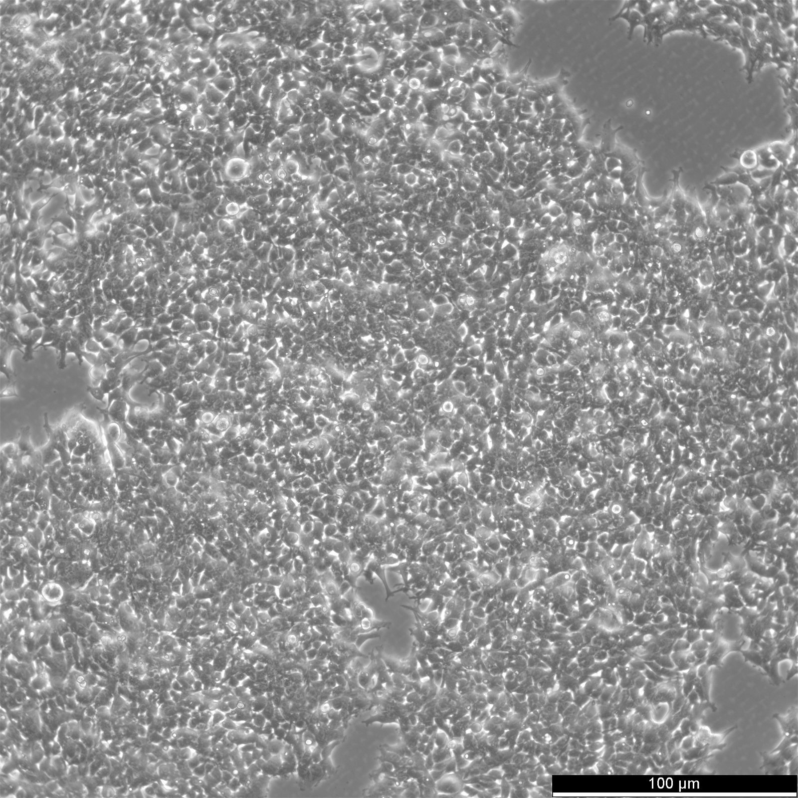
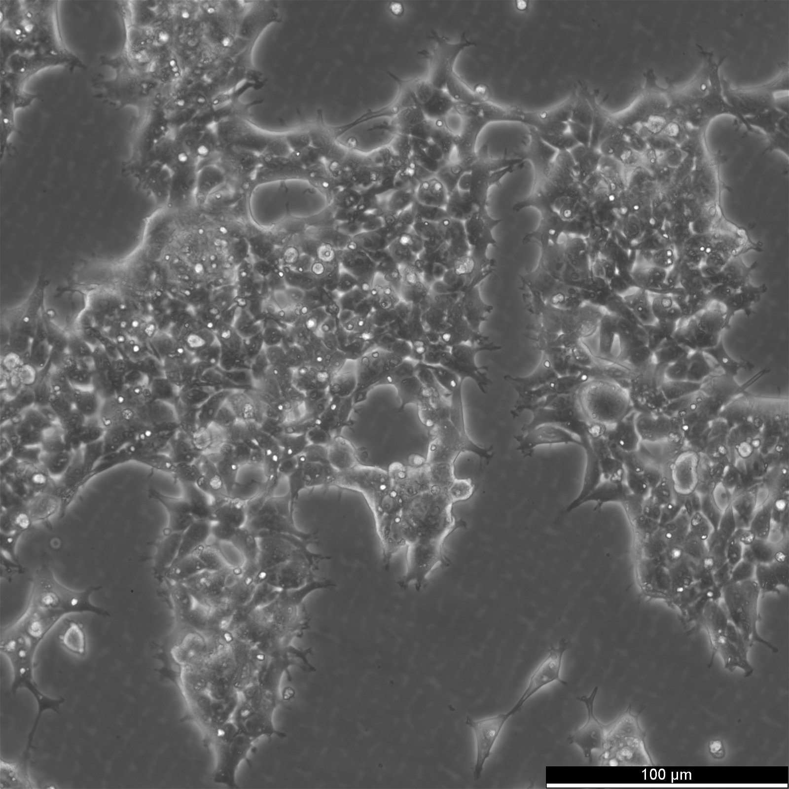
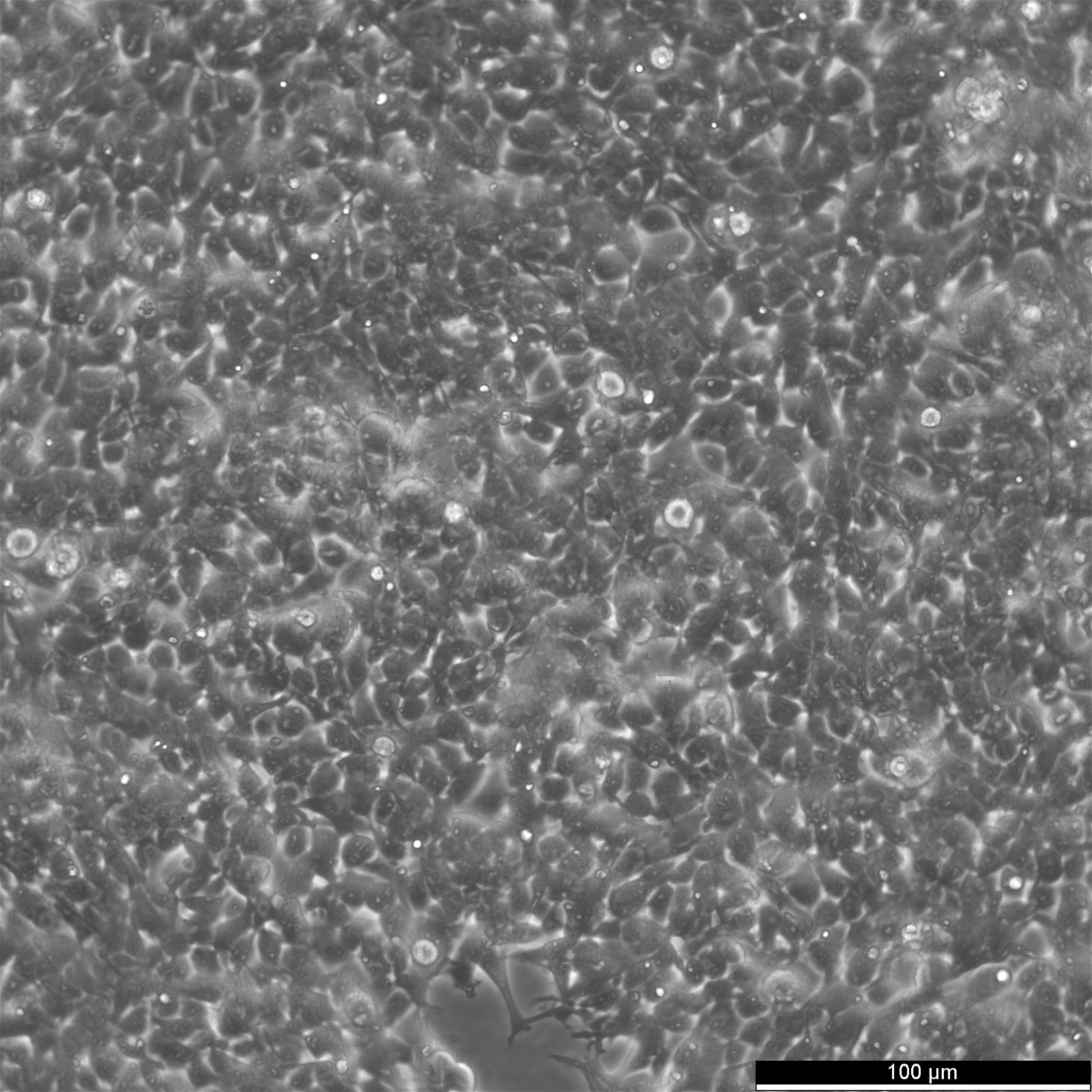
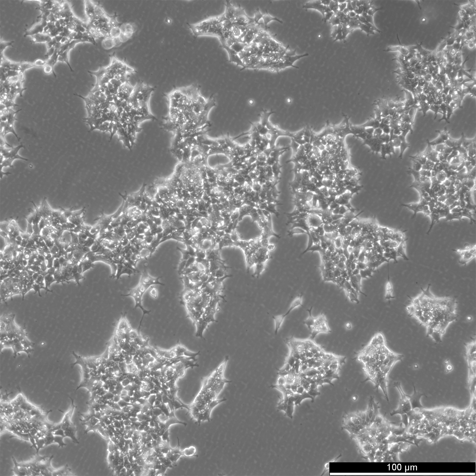
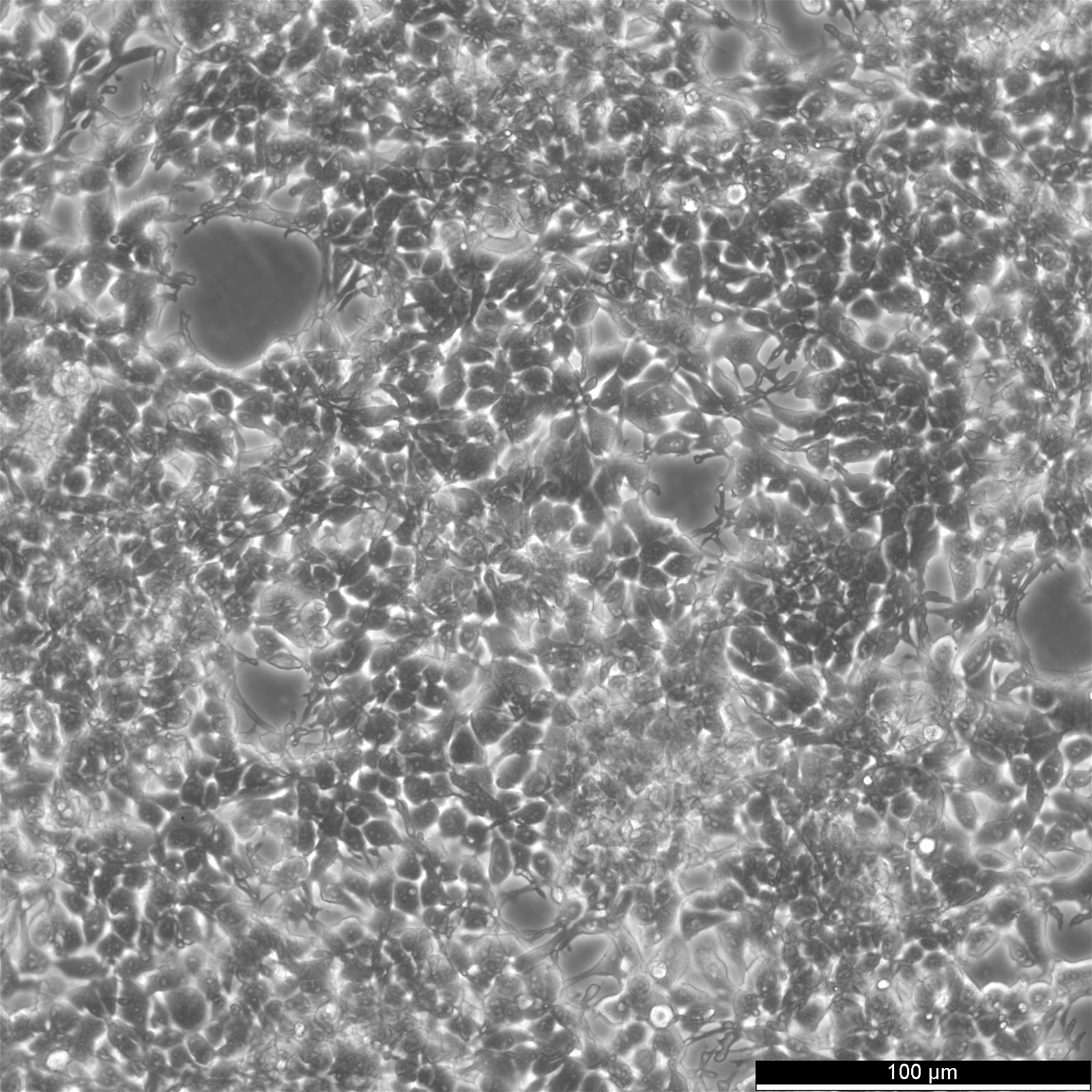
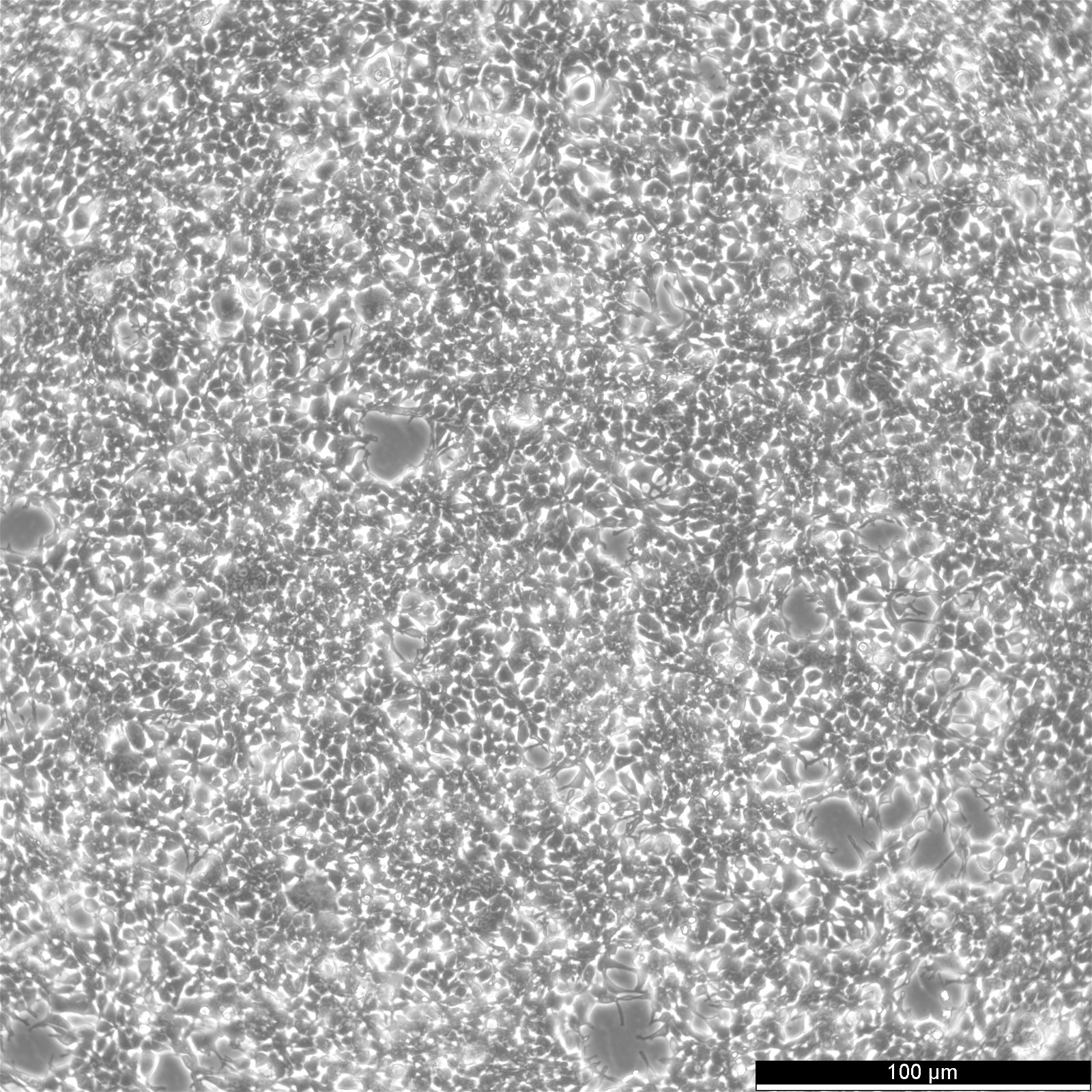
| Description | The AAV-293 cell line is a permanent line established from primary embryonic human kidney transformed with human adenovirus type 5 DNA. The genes encoded by the E1 region of adenovirus (E1a and E1b) are expressed in these cells and participate in transactivation of viral promoters, allowing these cells to produce high levels of protein. AAV-293 is derived from the parental 293 cell line, through cloning and multiple rounds of testing, AAV-293 is specifically selected for a high level of AAV production in a helper-free system. It offers several advantages over the regular 293 cells: Larger cell surface area resulting higher transfection and better yield of AAV. The advantages are a flattened morphology, firm attachment to culture plate, and the cells are ideal for large-scale culture and AAV production. Adeno-associated virus (AAV) belongs to the family of Parvoviridae, a group of viruses among the smallest of single-stranded and non-enveloped DNA viruses. There are nine different AAV serotypes reported to date. AAV can infect both dividing and non-dividing cells and can be maintained in the human host cell, creating the potential for long-term gene transfer. Recombinant AAV-2 is the most common serotype used in gene delivery, and can be produced at high titers with a helper virus or AAV-293 cells. |
|---|---|
| Organism | Human |
| Tissue | Embryonic kidney |
| Synonyms | AAV293 |
Characteristics
| Age | Fetus |
|---|---|
| Gender | Female |
| Morphology | Epithelial |
| Growth properties | Adherent |
Identifiers / Biosafety / Citation
| Citation | AAV-293 (Cytion catalog number 305127) |
|---|---|
| Biosafety level | 1 |
Expression / Mutation
Handling
| Culture Medium | DMEM, w: 4.5 g/L Glucose, w: 4 mM L-Glutamine, w: 1.5 g/L NaHCO3, w: 1.0 mM Sodium pyruvate (Cytion article number 820300a) |
|---|---|
| Medium supplements | Supplement the medium with 10% FBS, 0.1 mM NEAA |
| Passaging solution | Accutase |
| Subculturing | Remove the old medium from the adherent cells and wash them with PBS that lacks calcium and magnesium. For T25 flasks, use 3-5 ml of PBS, and for T75 flasks, use 5-10 ml. Then, cover the cells completely with Accutase, using 1-2 ml for T25 flasks and 2.5 ml for T75 flasks. Let the cells incubate at room temperature for 8-10 minutes to detach them. After incubation, gently mix the cells with 10 ml of medium to resuspend them, then centrifuge at 300xg for 3 minutes. Discard the supernatant, resuspend the cells in fresh medium, and transfer them into new flasks that already contain fresh medium. |
| Split ratio | 1:3 to 1:5 |
| Fluid renewal | 2 to 3 times per week |
| Freeze medium | CM-1 (Cytion catalog number 800100) or CM-ACF (Cytion catalog number 806100) |
| Handling of cryopreserved cultures | AAV-293 cells are shipped in a deep-frozen state on dry ice. Upon receipt, confirm that the vial remains frozen. For storage, place the cryovial immediately at temperatures below -150 degrees. If you plan to culture the cells immediately, swiftly thaw the vial by shaking it in a 37 degrees water bath with clean water and an antimicrobial agent for 40-60 seconds. Remove the vial once a small ice clump persists, ensuring it remains cold. Proceed with all subsequent steps under aseptic conditions. In a sterile flow hood, disinfect the cryovial with 70% ethanol. Then, gently open the vial and transfer the cell suspension into a 15 ml centrifuge tube pre-filled with 8 ml of room temperature culture medium. Gently mix the cells. For cell separation, centrifuge at 300 x g for 3 minutes and dispose of the supernatant. Skipping centrifugation is optional, although any residual freezing medium should be removed after 24 hours. Resuspend the pellet gently in 10 ml of fresh culture medium and divide between two T25 culture flasks. Follow the subculture protocol for subsequent steps. |
Quality control / Genetic profile / HLA
| Sterility | Mycoplasma contamination is rigorously excluded using both PCR-based assays and luminescence-based mycoplasma detection methods. To ensure there is no bacterial, fungal, or yeast contamination, cell cultures are subjected to daily visual inspections. |
|---|
