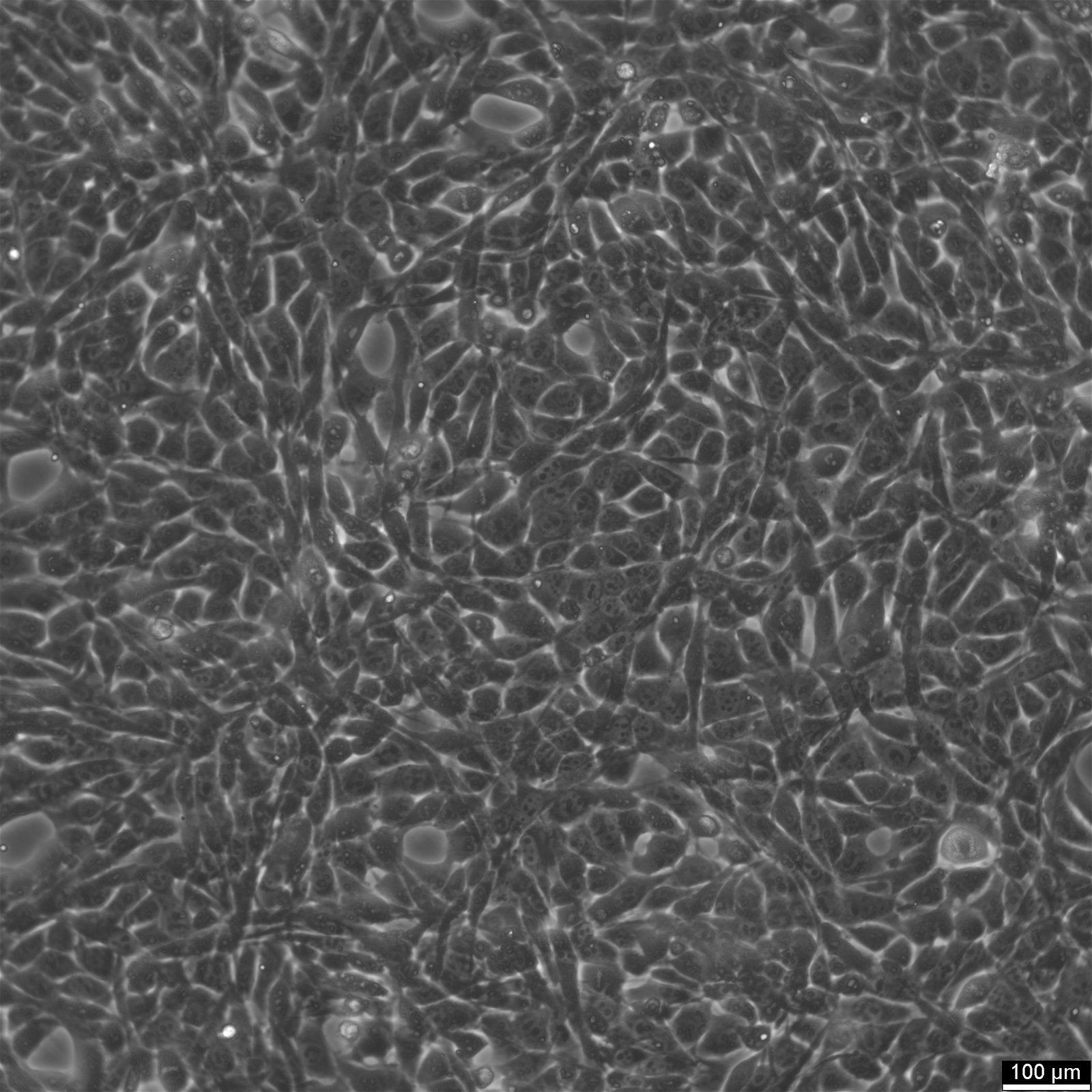
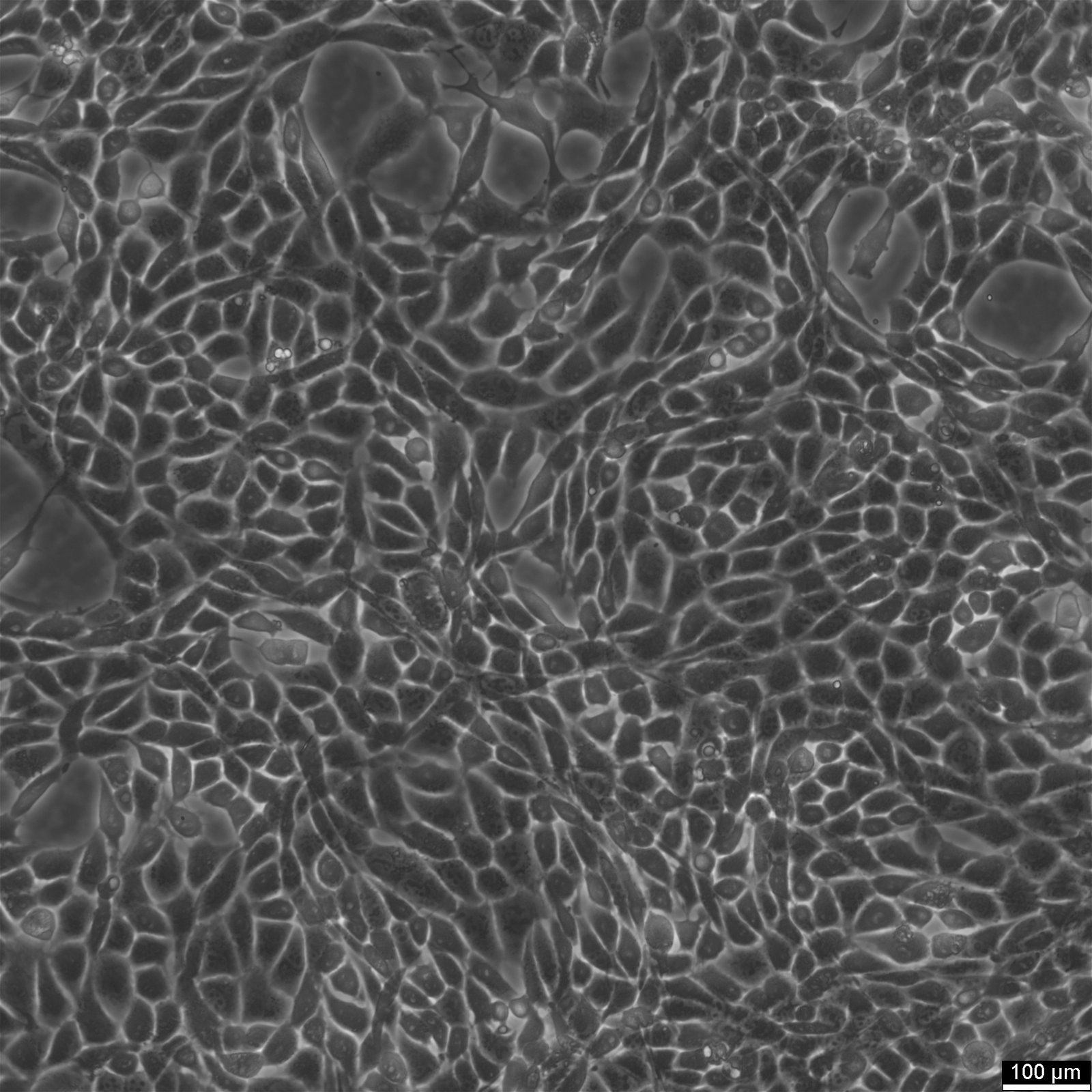
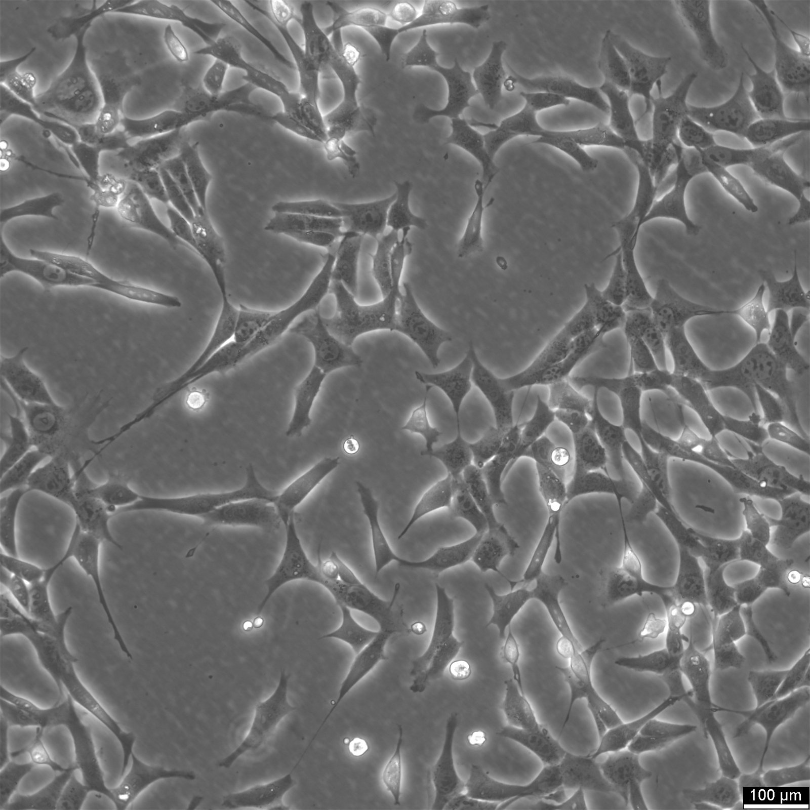
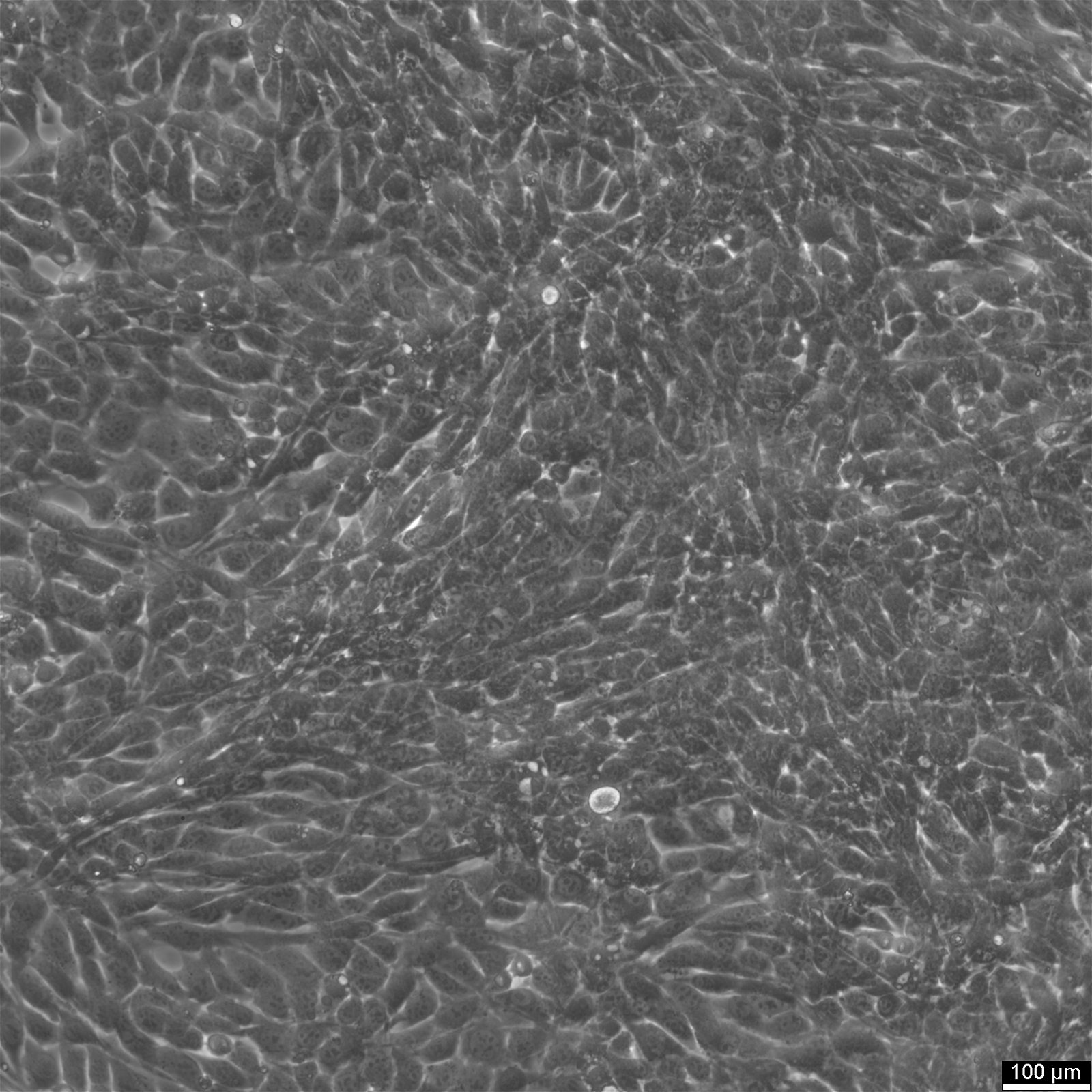
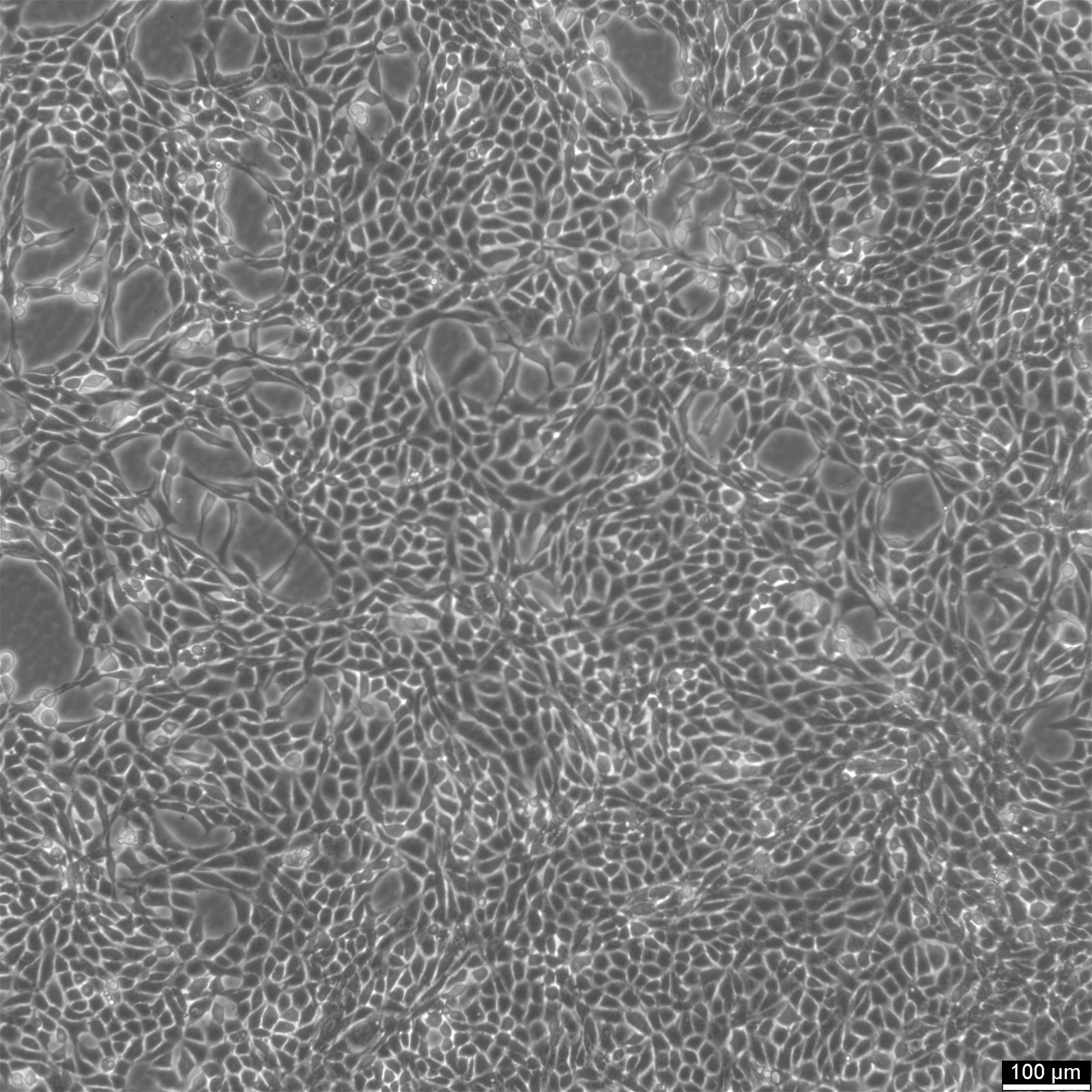
GC-1 spg


Product number:
300375
General information
Description | GC-1 spg Cells: A Breakthrough in 3D Cell Culture Derived from Mus musculus Testis GC-1 SPG cells are an extraordinary addition to the field of biological science, specifically in animal cell research. This innovative immortalized cell line originates from the testis of Mus musculus, a transgenic mouse engineered with the SV40 early region. Notably, GC-1 spg cells closely resemble spermatocytes, a critical male germ cell type, making them a precious tool for investigations in reproductive biology. Developed through the immortalization process facilitated by the SV40 large T antigen, GC-1 spg cells have demonstrated exceptional stability and viability. For 2.5 years, these cells have been cultured for an impressive 90 generations, showcasing their remarkable longevity and suitability for long-term studies. Distinct cell types contributing to the seminiferous tubule in the mouse testis have been immortalized and cultivated, resulting in a diverse collection of cell lines. This includes 16 peritubular cells, 22 Leydig cells, 8 Sertoli cells, and one germ cell line, all of which have shown exceptional growth and consistency throughout the culture period. Immortalized peritubular cells can be readily identified by their spindle-like morphology and their elevated expression of alkaline phosphatase and desmin, an intermediary filament. These cells exhibit substantial collagen production, highlighting their phenotypic characteristics. Leydig cells, another critical component of the seminiferous tubule, can be easily recognized by lipid droplets within their cytoplasm. Moreover, they exhibit heightened activity of the enzyme 3-beta-hydroxysteroid dehydrogenase and, in some instances, express LH receptors. These distinctive features make Leydig cells distinguishable and instrumental in various experimental setups. When cultured at high density, Sertoli cells showcase their typical in vivo columnar appearance. These immortalized Sertoli cells demonstrate an indented nucleus and cytoplasmic phagosomes, accurately reflecting their native characteristics. Notably, some Sertoli cell lines express FSH receptors, broadening the scope of research possibilities. The establishment of the GC-1 spg germ cell line represents a significant achievement. These cells occupy a crucial developmental stage, bridging the gap between spermatogonia type B and primary spermatocytes. Their distinct morphological and molecular features, observed through phase contrast and electron microscopy, allow for precise identification. Additionally, GC-1 spg cells express the testicular cytochrome ct and lactate dehydrogenase-C4 isozyme, confirming their resemblance to in vivo counterparts. Given their origin from the Mus musculus testis and their striking similarity to spermatocytes, GC-1 spg cells hold immense potential for various applications, with a particular focus on 3D cell culture. Researchers can utilize these cells to study the intricate processes involved in male germ cell development, uncovering valuable insights into reproductive biology and fertility. GC-1 spg cells represent a groundbreaking resource in the field of biological science. Derived from the testis of transgenic Mus musculus, these immortalized cells closely resemble spermatocytes, enabling researchers to delve into the complexities of male germ cell development. With their unique characteristics and robust culturing capabilities, GC-1 spg cells pave the way for groundbreaking discoveries in 3D cell culture and reproductive biology. |
---|---|
Organism | Mouse |
Tissue | Testis |
Applications | 3D cell culture |
Synonyms | GC-1spg, GC-1, GC1-SPG |
Characteristics
Age | 10 days |
---|---|
Gender | Male |
Morphology | Epithelial |
Cell type | Spermatocyte |
Growth properties | Adherent |
Identifiers / Biosafety / Citation
Citation | GC-1 spg (CLS catalog number 300375) |
---|---|
Biosafety level | 2 |
Expression / Mutation
Viruses | Transformant: Simian virus 40 (SV40) T antigen |
---|
Handling
Culture Medium | DMEM |
---|---|
Medium supplements | 10% FBS, w: 4.5 g/L Glucose, w: 4 mM L-Glutamine, w: 1.5 g/L NaHCO3, w: 1.0 mM Sodium pyruvate |
Passaging solution | Accutase |
Freeze medium | CM-1 (CLS catalog number 800100) or CM-ACF (CLS catalog number 806100) |
Handling of cryopreserved cultures | The cells come deep-frozen shipped on dry ice. Please make sure that the vial is still frozen. If immediate culturing is not intended, the cryovial must be stored below -150 degree Celsius after arrival. If immediate culturing is intended, please follow the below instructions: Quickly thaw by rapid agitation in a 37 degree Celsius water bath within 40-60 seconds. The water bath should have clean water containing an antimicrobial agent. As soon as the sample has thawed, remove the cryovial from the water bath. A small ice clump should still remain and the vial should still be cold. From now on, all operations should be carried out under aseptic conditions. Transfer the cryovial to a sterile flow cabinet and wipe with 70% alcohol. Carefully open the vial and transfer the cell suspension into a 15 ml centrifuge tube containing 8 ml of culture medium (room temperature). Resuspend the cells carefully. Centrifuge at 300 x g for 3 min and discard the supernatant. The centrifugation step may be omitted, but in this case the remains of the freeze medium have to be removed 24 hours later. Resuspend the cells carefully in 10 ml fresh cell culture medium and transfer them into two T25 cell culture flasks. All further steps are described in the subculture section. |
Handling of proliferating cultures | One or two cell culture flasks come filled with cell culture medium. Collect the entire medium in 1 or 2 x 50 ml centrifuge tubes, respectively. Carefully add 5 ml of cell culture medium to each T25 cell culture flask. Control the cell morphology and confluency under the microscope. Incubate at 37 degree Celsius for a minimum of 24 hours. Spin down the collected medium at 300 x g for 3 minutes to collect the cells which may have detached during transit. If a cell pellet is visible, resuspend the cells in 5 ml of cell culture medium and transfer to a T25 cell culture flask. Incubate at 37 degree Celsius for a minimum of 24 hours. |
Quality control / Genetic profile / HLA

Contamination-free cells
To identify mycoplasma contaminations we perform PCR-based and luminescence-based mycoplasma assays. We further determine any bacterial or fungal contamination through our standardized manufacturing processes.

Custom projects
Besides genomic DNA, RNA, cell pellets, and cell lysates, we can offer large quantities of assay-ready cells, plated cells in multiple formats, and frozen or growing cells. Contact us to receive a quote.

Authenticated cells
Each manufactured batch of cell lines* is authenticated via STR analysis. Contact us if you require a publication-ready STR report for your cells (*human, hamster, mouse, rat, and dog cells).

HLA alleles
HLA characterization is available from more than 200 cell lines. HLA class I -A, B, C, and Class II HLA-DPA1, -DPB1, -DQA1, DQB1, and DRB1 alleles were obtained by next-generation sequencing methodologies (NGS) for class I and class II alleles.