P19 Cells
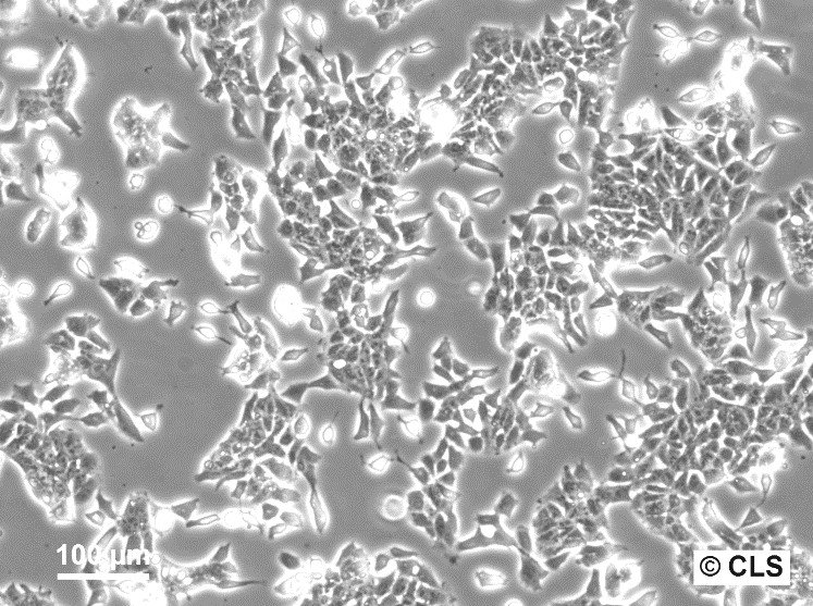
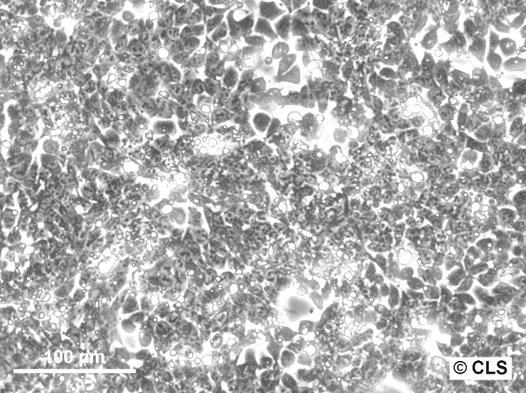
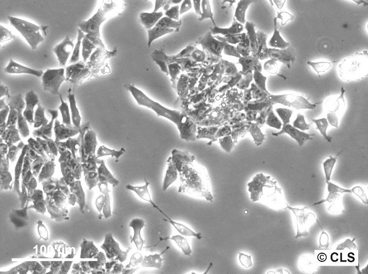
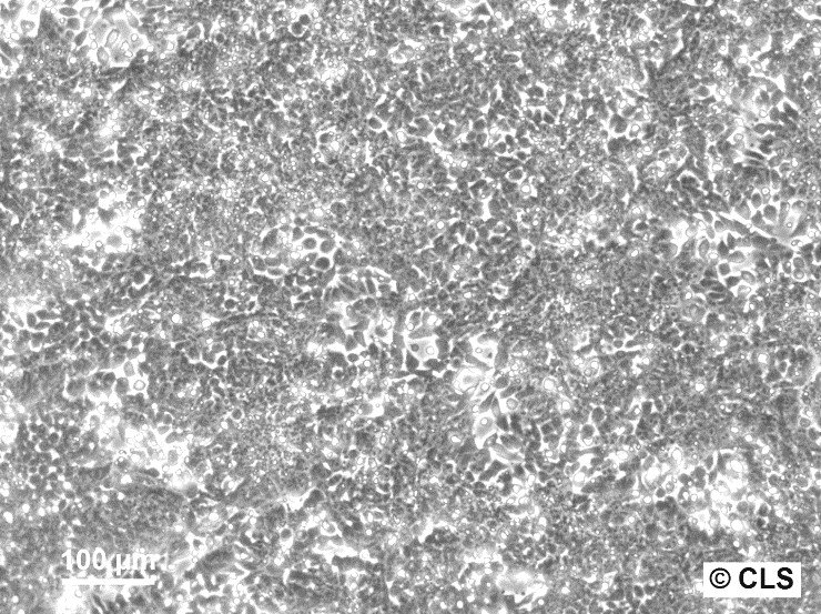


General information
| Description | The P19 cell line, a type of pluripotent embryonal carcinoma, was initially obtained from a teratocarcinoma in a C3H/He strain mouse. This epithelial-like cell line exhibits the capability to clone at high proficiency when grown in a medium infused with 0.1mM β-mercaptoethanol. A notable feature of P19 cells is their adaptability to differentiate into neuronal and glial cells when exposed to retinoic acid. Simultaneously, they have the potential to transform into cardiac and skeletal muscle when exposed to dimethyl sulfoxide (DMSO). When subjected to both retinoic acid and DMSO, they predominantly show characteristics of retinoic acid-induced differentiation. The P19 cell line has its origin in the mouse (Mus musculus) and belongs to the broad classification of Eukaryota, Animalia, Metazoa, Chordata, Vertebrata, and Tetrapod. The cells embody the morphology of an epithelial tissue type derived from the embryo and are associated with the disease teratocarcinoma. They are primarily utilized in 3D cell culture applications within the product category of animal cells. While cancer cells pose a significant health threat due to their rapid and aggressive growth, they also offer an invaluable resource for researchers studying cancer cell development and seeking more targeted treatments. In 1982, the P19 cell line was created when a 7.5-day mouse embryo was transplanted into a testis to induce tumour growth by McBurney and Rogers. They successfully isolated cell cultures from the primary tumour containing undifferentiated stem cells, termed embryonal carcinoma P19 cells. These cells demonstrated rapid growth without the need for feeder cells and were easy to maintain. Subsequent injection into blastocysts of another mouse strain confirmed the multipotency of P19 cells, as tissues from all three germ layers grew in the recipient mouse. Several subtype cell lines have been derived from the original P19 cells, including P19S18, P19D3, P19RAC65, and P19C16. Each of these subtypes possesses unique differentiation capabilities into neuronal cells or muscle cells when treated with retinoic acid or DMSO, respectively. More recent studies have generated cell lines derived from differentiated P19 cells, which, owing to the pluripotency of P19 cells, can transform into ectoderm, mesoderm, and endoderm-like cells. P19 cells are known for their sustained growth in serum-supplemented media. Their differentiation can be effectively controlled using nontoxic drugs such as retinoic acid, leading to the development of neurons, astroglia, and microglia. On the other hand, aggregates of P19 cells exposed to DMSO differentiate into endodermal and mesodermal derivatives, including cardiac and skeletal muscle. P19 cells are also amenable to transfection with DNA encoding recombinant genes, and stable lines expressing these genes can be conveniently isolated. This malleability and versatility make P19 cells an excellent resource for exploring the molecular mechanisms that govern the developmental decisions of differentiating pluripotent cells. |
|---|---|
| Organism | Mouse |
| Tissue | Testis |
| Disease | Teratocarcinoma |
| Synonyms | P-19 |
Characteristics
| Gender | Male |
|---|---|
| Morphology | Fibroblast-like |
| Growth properties | Adherent |
Identifiers / Biosafety / Citation
| Citation | P19 (Cytion catalog number 400416) |
|---|---|
| Biosafety level | 1 |
| Depositor | Burney |
Expression / Mutation
| Karyotype | n = 40, xY |
|---|
Handling
| Culture Medium | DMEM:Ham's F12, w: 3.1 g/L Glucose, w: 1.6 mM L-Glutamine, w: 15 mM HEPES, w: 1.0 mM Sodium pyruvate, w: 1.2 g/L NaHCO3 (Cytion article number 820400a) |
|---|---|
| Medium supplements | Supplement the medium with 10% FBS |
| Passaging solution | Accutase |
| Subculturing | Remove medium and rinse the adherent cells using PBS without calcium and magnesium (3-5 ml PBS for T25, 5-10ml for T75 cell culture flasks). Add TrypleExpress (1-2ml per T25, 2.5ml per T75 cell culture flask), the cell sheet must be covered completely. Incubate at 37 degree Celsius for 10 minutes. Carefully resuspend the cells, the addition of medium is optional but not necessary, and dispense into new flasks which contain fresh medium. Do not allow the cells to remain confluent. Subculture at least every 48 hours. |
| Split ratio | A ratio of 1:10 is recommended |
| Seeding density | Subculture at least every 48 hours |
| Fluid renewal | Every 2 days |
| Freeze medium | CM-1 (Cytion catalog number 800100) or CM-ACF (Cytion catalog number 806100) |
| Handling of cryopreserved cultures | P19 cells are shipped in a deep-frozen state on dry ice. Upon receipt, confirm that the vial remains frozen. For storage, place the cryovial immediately at temperatures below -150 degrees. If you plan to culture the cells immediately, swiftly thaw the vial by shaking it in a 37 degrees water bath with clean water and an antimicrobial agent for 40-60 seconds. Remove the vial once a small ice clump persists, ensuring it remains cold. Proceed with all subsequent steps under aseptic conditions. In a sterile flow hood, disinfect the cryovial with 70% ethanol. Then, gently open the vial and transfer the cell suspension into a 15 ml centrifuge tube pre-filled with 8 ml of room temperature culture medium. Gently mix the cells. For cell separation, centrifuge at 300 x g for 3 minutes and dispose of the supernatant. Skipping centrifugation is optional, although any residual freezing medium should be removed after 24 hours. Resuspend the pellet gently in 10 ml of fresh culture medium and divide between two T25 culture flasks. Follow the subculture protocol for subsequent steps. |
Quality control / Genetic profile / HLA
| Sterility | Mycoplasma contamination is rigorously excluded using both PCR-based assays and luminescence-based mycoplasma detection methods. To ensure there is no bacterial, fungal, or yeast contamination, cell cultures are subjected to daily visual inspections. |
|---|---|
| STR profile |
Amelogenin: x,x
|
Required products
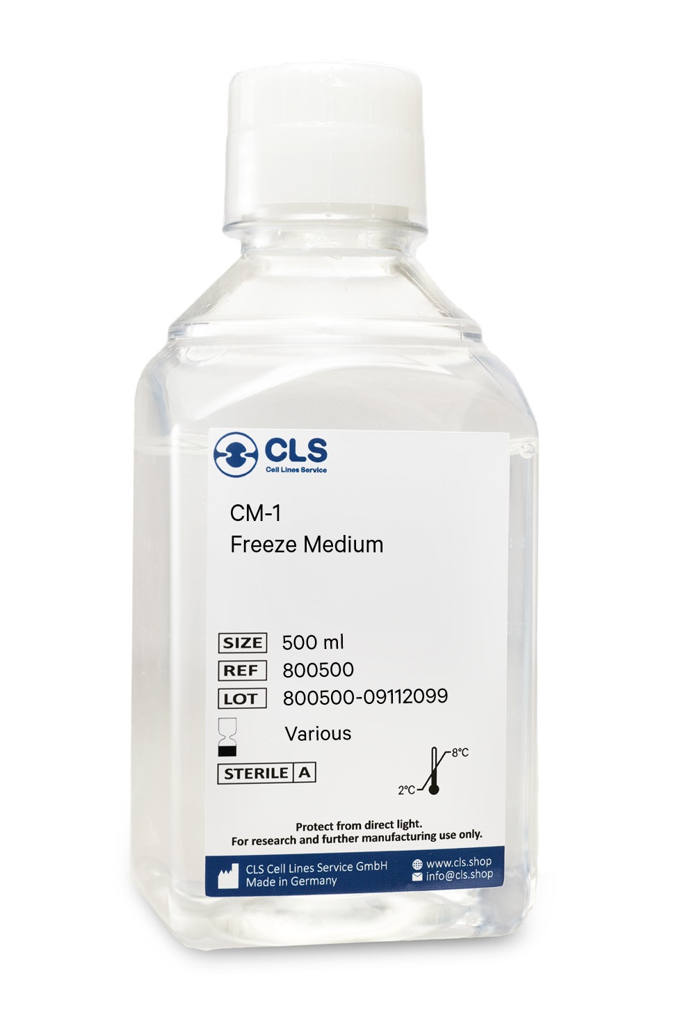
In biological research, the cryopreservation of mammalian cells is an invaluable tool. Successful preservation of cells is a top priority given that losing a cell line to contamination or improper storage conditions leads to lost time and money, ultimately delaying research results. Once the cells have been transferred from a cell growth medium to a freezing medium, the cells are typically frozen at a regulated rate and stored in liquid nitrogen vapor or at below -130°C in a mechanical deep freezer. The freeze medium CM-1 enables cryopreservation of cells at below -130°C (or in liquid nitrogen), essentially eliminating the need for an additional, costly ultralow freezer and eliminating time-consuming and demanding controlled rate freezing processes. Simply collect the cells, aspirate the growth medium, resuspend in CM-1, transfer to a cryovial, and store the vial at below -130 °C.
Long shelf-life
CM-1 is a serum-containing, ready-to-use cryopreservation medium that can be stored in the refrigerator for up to one year.
Trusted by hundreds of researchers
Our advanced cell freezing medium CM-1 is a market-leading product in Germany and Europe and is distinguished by numerous publications involving hundreds of different cell lines worldwide. We tested it with more than 1000 cell lines from our proprietary cell bank.
Optimized ingredients
CM-1 does contain serum products. Serum-containing cryopreservation mediums optimally protect the cells whilst being frozen and have the advantage of high recovery rates. As CM-1 has been tested with a multitude of cell lines, you can rest assured that your cells always recover well.
Contains FBS, DMSO, glucose, salts
Buffering capacity pH = 7.2 to 7.6
Applications & Validation
The cells preserved in our CM-1 freeze medium can be used for cell counting, viability and cryopreservation, cell culture, mammalian cell culture, gene expression analysis and genotyping, in vitro transcription, and polymerase chain reactions. Each batch's efficacy is evaluated using CHO-K1 cells. Each batch is tested for pH, osmolality, sterility, and endotoxins to ensure high quality.
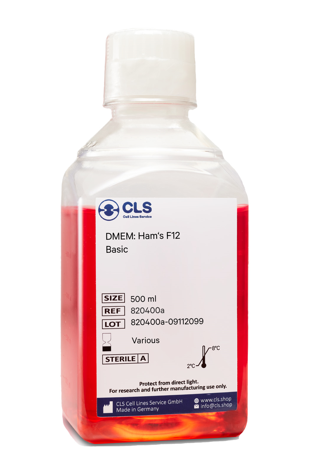
This unique formulation combines Dulbecco's Modified Eagle Medium (DMEM) and Ham's F-12 (Ham's Nutrient Mixture F-12) in a precise 1:1 ratio. The addition of L-glutamine further enhances its composition.
DMEM, derived from Eagle's Minimal Essential Medium (EMEM), offers an increased concentration of amino acids and vitamins compared to its predecessor. In contrast, Ham's F-12 is based on Ham's F-10 medium, providing a complementary set of essential components.
To support optimal cell growth, it is common practice to supplement DMEM:Ham's F12 with FBS at a typical concentration of 5-10%. This addition is necessary as the medium lacks growth hormones, lipids, and proteins crucial for cellular development.
DMEM:Ham's F12 incorporates a pH buffer system and is often supplemented with phenol red, a pH indicator. Cultured cells in DMEM:Ham's F12, or any medium utilizing the bicarbonate buffer system, require a controlled CO2 environment of 5-10% to maintain appropriate pH levels. Phenol red enables monitoring of pH changes from 6.2 (yellow) to 8.2 (red).
Quality Control
pH = 7.2 +/
- 0.02 at 20-25°C.
Each lot has been tested for sterility and absence of mycoplasma and bacteria.
Maintenance
Keep refrigerated at +2°C to +8°C in the dark. Freezing as well as warming up to +37°C minimize the quality of the product.
Do not heat the medium to more than 37°C or use uncontrollable sources of heat (e.g., microwave appliances).
If only a part of the medium is to be used, remove this amount from the bottle and warm it up at room temperature.
Shelf life for any medium except for the basic medium is 8 weeks from the date of manufacture.
Composition
Components
mg/L
Inorganic Salts
Calcium chloride x 2H2O
154,45
Iron(III)-nitrate x 9H2O
0,05
Iron(II)-sulfate x 7H2O
0,42
Potassium chloride
311,83
Copper(II)-sulfate x 5H2O
0.001
Magnesium chloride anhydrous
28,57
Magnesium sulfate
48,85
Sodium chloride
6,999.50
Sodium dihydrogen phosphate anhydrous
54,35
di-Sodium hydrogen phosphate
70,98
Zinc sulfate x 7H2O
0,43
Other Components
D(+)-Glucose anhydrous
3,151.00
HEPES
3,574.50
Hypoxanthine
2,04
Linoleic acid
0,04
DL-68-Lipoic acid
0.103
Sodium pyruvate
110,00
Phenol red
8,10
Putrescin x 2HCl
0.081
Thymidine
0,36
Amino Acids
L-Alanine
4,45
L-Arginine x HCl
147,35
L-Asparagine x H2O
7,50
L-Aspartic acid
6,65
L-Cystine x 2HCl
31,29
L-Cysteine x HCl x H2O
17,56
L-Glutamine
365,00
L-Glutamic acid
7,35
Glycine
18,75
L-Histidine x HCl x H2O
31,48
L-Isoleucine
54,37
L-Leucine
58,96
L-Lysine x HCl
91,37
L-Methionine
17,24
L-Phenylalanine
35,48
L-Proline
17,27
L-Serine
26,25
L-Threonine
53,55
L-Tryptophan
9,02
L-Tyrosine x 2Na x 2H2O
55,81
L-Valine
52,66
Vitamins
D-(+)-Biotine
0,004
D-Calcium pantothenate
2,12
Choline chloride
8,98
Folic acid
2,66
myo-Inositol
12,51
Nicotinamide
2,02
Pyridoxine x HCl
2,03
Riboflavin
0,22
Thiamine x HCl
2,17
Vitamine B12
0,68
NaHCO3
1,200.00
- A Gentle Alternative to Trypsin
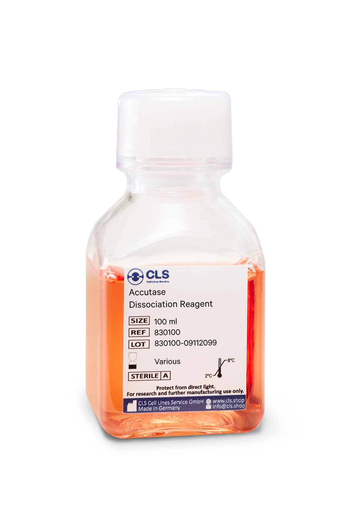
Accutase is a cell detachment solution that is revolutionizing the cell culture industry. It is a mix of proteolytic and collagenolytic enzymes that mimics the action of trypsin and collagenase. Unlike trypsin, Accutase does not contain any mammalian or bacterial components and is much gentler on cells, making it an ideal solution for the routine detachment of cells from standard tissue culture plasticware and adhesion coated plasticware. In this blog post, we will explore the benefits and uses of Accutase and how it is changing the game in cell culture.
Advantages of Accutase
Accutase has several advantages over traditional trypsin solutions. Firstly, it can be used whenever gentle and efficient detachment of any adherent cell line is needed, making it a direct replacement for trypsin. Secondly, Accutase works extremely well on embryonic and neuronal stem cells, and it has been shown to maintain the viability of these cells after passaging. Thirdly, Accutase preserves most epitopes for subsequent flow cytometry analysis, making it ideal for cell surface marker analysis.
Additionally, Accutase does not need to be neutralized when passaging adherent cells. The addition of more media after the cells are split dilutes Accutase so it is no longer able to detach cells. This eliminates the need for an inactivation step and saves time for cell culture technicians. Finally, Accutase does not need to be aliquoted, and a bottle is stable in the refrigerator for 2 months.
Applications of Accutase
Accutase is a direct replacement for trypsin solution and can be used for the passaging of cell lines. Additionally, Accutase performs well when detaching cells for the analysis of many cell surface markers using flow cytometry and for cell sorting. Other downstream applications of Accutase treatment include analysis of cell surface markers, virus growth assay, cell proliferation, tumor cell migration assays, routine cell passage, production scale-up (bioreactor), and flow cytometry.
Composition of Accutase
Accutase contains no mammalian or bacterial components and is a natural enzyme mixture with proteolytic and collagenolytic enzyme activity. It is formulated at a much lower concentration than trypsin and collagenase, making it less toxic and gentler, but just as effective.
Efficiency of Accutase
Accutase has been shown to be efficient in detaching primary and stem cells and maintaining high cell viability compared to animal origin enzymes such as trypsin. 100% of cells are recovered after 10 minutes, and there is no harm in leaving cells in Accutase for up to 45 minutes, thanks to autodigestion of Accutase.
In summary
In conclusion, Accutase is a powerful solution that is changing the game in cell culture. With its gentle nature, efficiency, and versatility, Accutase is the ideal alternative to trypsin. If you are looking for a reliable and efficient solution for cell detachment, Accutase is the solution for you.
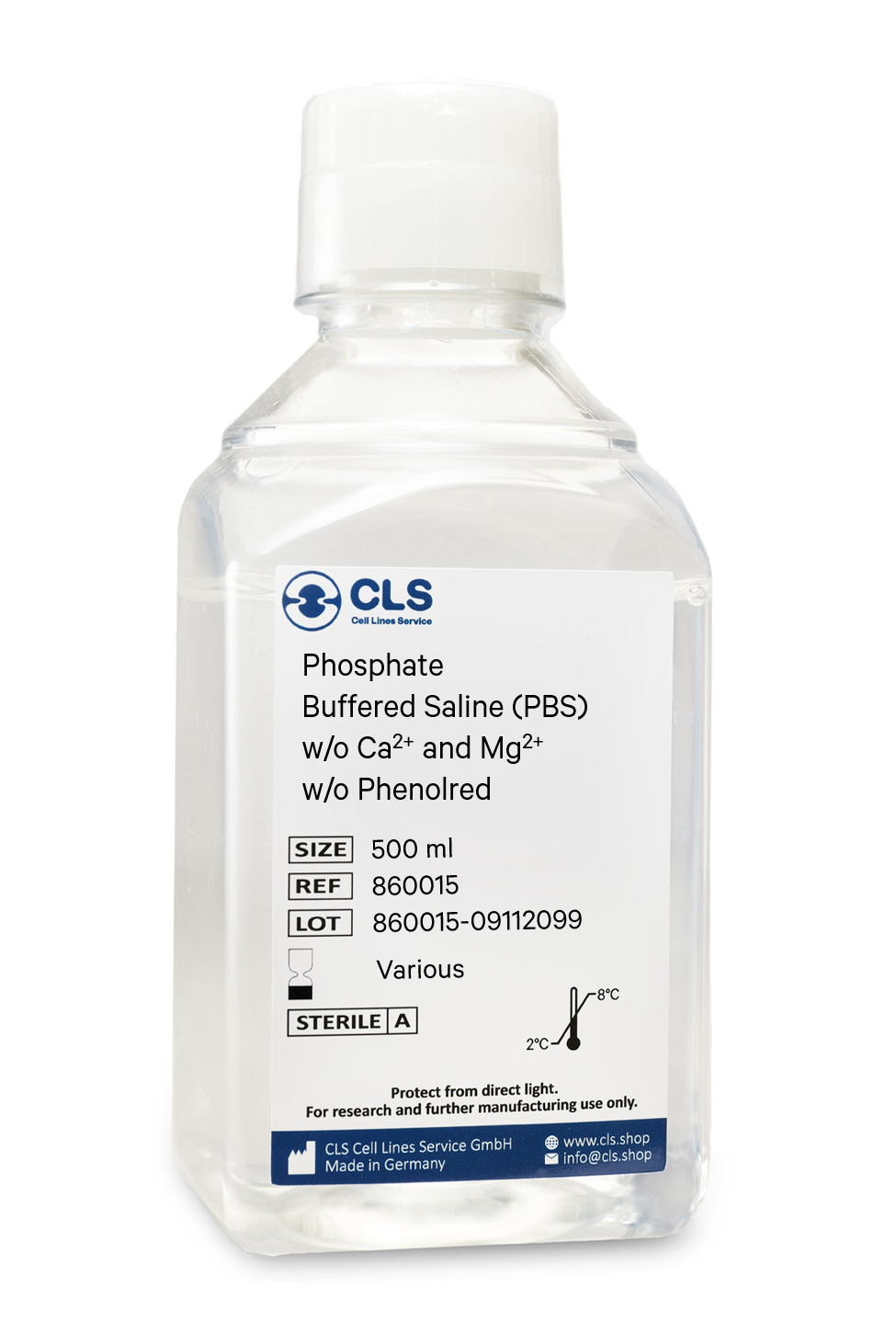
Phosphate-buffered saline (PBS) is a versatile buffer solution used in many biological and chemical applications, as well as tissue processing. Our PBS solution is formulated with high-quality ingredients to ensure a constant pH during experiments. The osmolarity and ion concentrations of our PBS solution are matched to those of the human body, making it isotonic and non-toxic to most cells.
Composition of our PBS Solution
Our PBS solution is a pH-adjusted blend of ultrapure-grade phosphate buffers and saline solutions. At a 1X working concentration, it contains 137 mM NaCl, 2.7 mM KCl, 8 mM Na2HPO4, and 2 mM KH2PO4. We have chosen this composition based on CSHL protocols and Molecular cloning by Sambrook, which are well-established standards in the research community.
Applications of our PBS Solution
Our PBS solution is ideal for a wide range of applications in biological research. Its isotonic and non-toxic properties make it perfect for substance dilution and cell container rinsing. Our PBS solution with EDTA can also be used to disengage attached and clumped cells. However, it is important to note that divalent metals such as zinc cannot be added to PBS as this may result in precipitation. In such cases, Good's buffers are recommended. Moreover, our PBS solution has been shown to be an acceptable alternative to viral transport medium for the transport and storage of RNA viruses, such as SARS-CoV-2.
Storage of our PBS Solution
Our PBS solution can be stored at room temperature, making it easy to use and access.
To sum up
In summary, our PBS solution is an essential component in many biological and chemical experiments. Its isotonic and non-toxic properties make it suitable for numerous applications, from cell culture to viral transport medium. By choosing our high-quality PBS solution, researchers can optimize their experiments and ensure accurate and reliable results.
Composition
Components
mg/L
Inorganic Salts
Potassium chloride
200,00
Potassium dihydrogen phosphate
200,00
Sodium chloride
8,000.00
di-Sodium hydrogen phosphate anhydrous
1,150.00

