
MX-1 Cell Line
MX-1 is a human breast adenocarcinoma cell line derived from a breast cancer xenograft model in athymic mice. The human breast adenocarcinoma cells were established from the breast tumor of a 29-year-old Caucasian female. MX1 cells are triple-negative breast cancer (TNBC) that are extensively utilized for research on drug resistance, cell signaling, drug response mechanism, etc.
This article provides information regarding the MX-1 cell line. Here are several vital points that will be discussed:
- Origin and General Information on MX-1 Cells
- MX-1 cell line: Culturing information
- MX-1 cells: Applications in Research
- Publications related to MX-1 cells
- MX-1 cell line Resources: Protocols, Videos & More
1. Origin and General Information on MX-1 Cells
Before working with this cell line, you should learn its nuts and bolts. We will review almost all the general information regarding the MX-1 cell line.
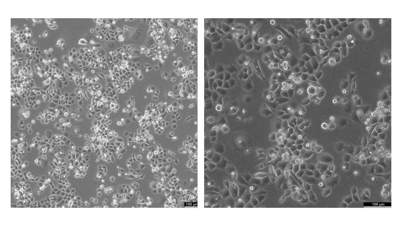
The immortal MX-1 cell line is a tumorigenic cell line established from a tumor xenograft. MX-1 cells are considered a type of triple-negative breast cancer (TNBC) [1]. Moreover, these cells are estrogen receptor and p53 negative, as they do not express these genes.
MX-1 cells - A Model System for Triple-Negative Breast Cancer
The culture conditions of the MX-1 cell line are not fussy. These cells can be easily propagated and used in various research laboratories for transfection studies. The transient transfection method commonly studies the underlying cellular mechanisms and gene expression patterns in this cell line [2, 3].
This cell line is a sound model system for breast carcinomas. It can be used to understand the molecular mechanisms behind disease progression and to screen and develop drugs to combat disease. More specifically, the human MX-1 breast adenocarcinoma cell line can be an alternative to TNBC cells as it is estrogen receptor-negative [4].
2. MX-1 cell line: Culturing information
Key Points for Culturing MX-1 Cells
|
Doubling Time: |
The population doubling time of INS-1 cells is approximately 44 hours. |
|
Adherent or in Suspension: |
INS-1 cells grow both in suspension and adherent form. |
|
Sub-cultivation Ratio: |
INS-1 cells are sub-cultured at a split ratio of 1:3. Briefly, suspended cells are collected. Adherent cells are rinsed with PBS and incubated with the Accutase solution. After detachment cells are added with fresh media. Afterwards, both suspended and adherent cells are centrifuged and collected. Cells are carefully resuspended and dispensed into the new flasks for growth. |
|
Growth Medium: |
RPM1 1640 is used to culture the INS-1rat insulinoma cell line. The media is supplemented with 10% heat-inactivated fetal bovine serum, 2.1 mM stable Glutamine, 10 mM HEPES, 2.0 g/L NaHCO3, and 1 mM sodium pyruvate. |
|
Growth Conditions: |
INS-1 cells are kept in a humidified incubator set at 37°C temperature and with a continuous 5% CO2 supply. |
|
Storage: |
INS-1 beta cells can be stored in the vapour phase of liquid nitrogen or at below -150°C temperature in an electric freezer for the long term. |
|
Freezing Process and Medium: |
CM-1 or CM-ACF media are used to freeze INS-1 cells through a slow freezing process. It allows only a 1°C drop in temperature per minute to protect cell viability. |
|
Thawing Process: |
Frozen INS-1 cells are thawed in a water bath pre-set at 37 degrees Celsius temperature for 40 to 60 seconds. After thawing, cells are added with fresh media and directly poured into a new flask for growth. After 24 hours media is replaced to eliminate freezing media components. |
|
Biosafety Level: |
Biosafety level 1 laboratory is required to culture INS-1 rat insulinoma cells. |
3. MX-1 cells: Applications in Research
This human breast adenocarcinoma MX-1 cell line is commonly used in breast cancer studies instead of cells such as MDA-MB-231 or MCF-7. Here are a few special applications of this cell line in cancer research.
- Drug resistance mechanisms
MX-1 cells are used to study the molecular factors mediating the development of resistance against breast cancer drugs. Studies have widely used MX-1 cells to establish drug-resistant models. A study published in 2021 reported the use of MX-1 cells to generate a doxorubicin-resistant breast cancer cell line. The developed cell model validated the ABC transporter ABCB1 and epithelial-mesenchymal transition (EMT) involvement in acquiring chemoresistance [5].
- Gene expression/cell signaling pathway
The MX-1 cell line can be transfected transiently to elucidate gene expression patterns and cell signaling pathways. Studies have used MX-1 cells for shRNA (short hairpin RNA) and non-coding RNA transfection to explore their effect on breast cancer cells proliferation and growth. Moreover, associated gene signaling pathways are elucidated [2, 6].
- Screening of potential inhibitors
MX-1 cells can be used for screening potential drugs against breast cancer as they mimic the cancer cell microenvironment. A research study has shown the therapeutic activity of Vinorelbine, a microtubule toxin, that triggered cell death and polyploidy in MX-1 cells [4].
4. Publications related to MX-1 cells
Table 1. Notable Publications with MX-1 Cells
|
Journal |
Year |
Study Title |
Cell Lines Used |
Key Findings |
|
Nature: Scientific Reports |
2021 |
Nongenotoxic ABCB1 activator tetraphenylphosphonium can contribute to doxorubicin resistance in MX-1 breast cancer cell line |
MX-1 |
Doxorubicin resistance MX-1 cell line was developed to study molecular mechanisms, i.e., epithelial-mesenchymal transition (EMT) and ABC transporter ABCB1. |
|
Cancer & Chemotherapy |
2019 |
MX-1 |
The anti-breast cancer potential of a microtubule toxin, Vinorelbine, was evaluated. |
|
|
Apoptosis |
2021 |
MX-1, MDA-MB-231 |
Estrogen receptor-negative MX-1 and MDA-MB-231 cell lines were used to study the molecular factors behind the cell death pathway activation. |
|
|
International Journal of Molecular Medicine |
2021 |
MCF‑7, MX‑1, MDA‑MB‑231 |
The radio-sensitization effect of Bosutinib was explored. The drug makes cells radiation sensitive by targeting eIF4G1 and other DNA damage response proteins. |
|
|
The International Journal of Biochemistry & Cell Biology |
2018 |
Mixomics analysis of breast cancer: Long non-coding RNA linc01561 acts as ceRNA involved in the progression of breast cancer |
MX-1 |
The role of linc01561 long non-coding RNA in breast cancer development was explored via transfection of MX-1 cells. |
5. MX-1 cell line Resources: Protocols, Videos & More
There are limited resources available regarding culturing and transfection methods, but we gathered as much information as possible for you.
Cell culture protocols
MX-1 cells are used in many transient transfection analysis studies. Here, we have listed a few resources to help you with the transfection protocols.
- MX-1 cells transfection: This publication describes the protocol for transfection with siRNA in MX-1 cells for developing a gene knock-out model.
- Mammalian cell transfection: This article has all the essential information about transfection methods used in mammalian cell lines.
We hope that this article has provided you with valuable knowledge on the MX-1 cell line and that you have gained a better understanding of how to culture, maintain, and utilize these cells in your research. If you're interested in working with the MX-1 cell line, don't hesitate to order from us to kickstart your research journey!
References
- Stefanski, C.D. and J.R. Prosperi, Combating CHK1 resistance in triple negative breast cancer: EGFR inhibition as potential combinational therapy. Cancer Drug Resistance, 2022. 5(1): p. 229.
- Vasiyani, H., et al., The analog of cGAMP, c-di-AMP, activates STING mediated cell death pathway in estrogen-receptor negative breast cancer cells. Apoptosis, 2021. 26: p. 293-306.
- Xiang, S., et al., Proteomic analysis of inhibitor of apoptosis protein‑like protein‑2 on breast cancer cell proliferation. Molecular Medicine Reports, 2022. 25(3): p. 1-11.
- Nakajima, H., C. Furukawa, and J. Magae, Vinorelbine, a Microtubule Toxin, Induces Apoptosis and Polyploidy in MX-1, a Human Triple-Negative Breast Cancer Cell Line. Gan to Kagaku ryoho. Cancer & Chemotherapy, 2019. 46(3): p. 447-451.
- Kubiliute, R., et al., Nongenotoxic ABCB1 activator tetraphenylphosphonium can contribute to doxorubicin resistance in MX-1 breast cancer cell line. Scientific reports, 2021. 11(1): p. 1-11.
- Jiang, R., et al., Mixomics analysis of breast cancer: Long non-coding RNA linc01561 acts as ceRNA involved in the progression of breast cancer. The International Journal of Biochemistry & Cell Biology, 2018. 102: p. 1-9.