
NIH-3T3 Cells: Advancing Fibroblast Studies and Applications of NIH-3T3
The NIH-3T3 cell line, established from the tissue of a 17-day-old Swiss Albino mouse embryo in 1962 by Howard Green and George Todaro at the New York University School of Medicine, has become a fundamental resource in biomedical research. Recognized for its high receptiveness to leukemia virus and sarcoma virus focus formation, NIH-3T3 cells serve as a critical tool for a plethora of scientific inquiries, including viral oncology studies, gene expression analysis, and exploration of cellular growth dynamics. The "3T3" nomenclature reflects the cell culture method, denoting a "3-day transfer" interval with an initial seeding density of 3 × 10^5 cells, highlighting the standardized conditions under which these cells were first cultured and expanded.
Diverse Morphologies and Applications of NIH-3T3 Cells
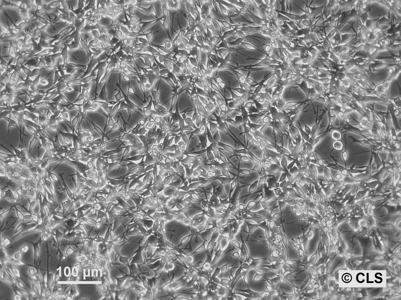
One of the hallmark characteristics of NIH-3T3 cells is their morphological adaptability, which varies significantly with culture confluency. At lower densities, these fibroblasts display a spindle-shaped, solitary cell structure, evolving into dense, swirling patterns as the population reaches confluence. With an average diameter of about 18 μm, NIH-3T3 cells offer a versatile model for in-depth cell biology studies, ranging from tissue repair mechanisms to the intricate pathways of cell cycle regulation.

Culturing information
Key Culturing Details:
Population Doubling Time: Roughly 20 hours.
Growth Type: Adherent cultures.
Seeding Density: Recommended: 3 to 4 x 10^4 cells/cm^2.
Growth medium: DMEM or Ham's F12, supplemented with 5% FBS and 2.5 mM L-glutamine.
Growth Conditions: Maintain at 37 °C in a humidified incubator with 5% CO2.
Storage: Keep at temperatures below -195 °C in the vapor phase of liquid nitrogen.
Freezing Method: Use CM-1 or CM-ACF medium; employ a slow freezing method (1°C temperature drop).
Thawing Protocol: rapid warming in a 37 °C water bath, followed by centrifugation to remove freezing medium, then resuspension in growth medium.
Biosafety Level: Culturing requires a biosafety level 1 setting.

Swiss Albino mouse in a laboratory.
Pros and Cons of Using NIH 3T3 Cells
Advantages
Transfection Efficiency: Known for their high transfection rates, NIH-3T3 cells are excellent for both transient and stable gene expression studies, accommodating a variety of transfection techniques.
Feeder Layer Utility: These cells often serve as a supportive feeder layer for co-cultures with cells like keratinocytes and stem cells, thanks to their release of growth factors that promote co-cultured cell growth.
Stem Cell Research: NIH-3T3 cells are a preferred choice in stem cell research for inducing pluripotency without genetic modification and providing a conducive environment for stem cell differentiation.
Culture stability: NIH-3T3 cells are known for their stability and low frequency of spontaneous transformation. However, under certain conditions or after exposure to specific oncogenes or mutagens, NIH-3T3 cells can undergo spontaneous transformation. This transformation can lead to the acquisition of cancerous properties such as uncontrolled growth, loss of contact inhibition, and the ability to form tumors when injected into susceptible hosts.
Disadvantages
Inconsistent Cell Size: The elongated, spindle-like morphology of NIH-3T3 cells can vary, complicating image analyses in assays.
Infection Susceptibility: These cells are prone to bacterial and mycoplasma infections if not maintained in stringent aseptic conditions, potentially impacting experimental integrity.
Research Applications of NIH-3T3 Cells
DNA Transfection Studies: NIH-3T3 cells' robustness makes them ideal for introducing and studying the function of various genes, demonstrated in research examining proteins like NAB2-STAT6 and their roles in cellular processes.
Cell-Based Assays: Their reliability extends to various assays, including viability, apoptosis, and focus formation assays, offering insights into cellular responses under different experimental conditions.
Cell Cycle Research: The cell line's straightforward cell cycle manipulation via serum levels makes it a potent model for studying cell cycle regulation and its aberrations in disease contexts.
Elevate Your Research with NIH-3T3 Cells
Highlighting Key Studies Involving the fibroblast cell line NIH 3T3
The NIH-3T3 cell line has been pivotal in numerous research projects, spanning various facets of cellular biology. Below are some significant studies utilizing these cells:
- Exploring the NAB2-STAT6 Fusion Protein: Published in Biochemical and Biophysical Research Communications, this study delves into how the NAB2-STAT6 fusion protein impacts NIH-3T3 cells, specifically its role in enhancing cell growth and migration through EGR-1 regulation.
- Investigating APOBEC3 and Murine Leukemia Virus: This research in Virology journal examines the hypermutation of AKV murine leukemia virus in NIH-3T3 cells expressing the mouse APOBEC3 gene.
- Evaluating Antimetastatic Potential of Epigenetic Drugs: In Oncotargets and Therapy, this study assesses the antimetastatic effects of hydralazine and valproic acid on RAS-transformed NIH-3T3 cells.
- Baicalein's Impact on NIH-3T3 Proliferation and Collagen Synthesis: This research utilizes NIH-3T3 cells to explore how Baicalein influences cell proliferation and collagen production through miR-9/insulin-like growth factor-1 axis modulation.
- Studying Riboflavin Depletion and Tumorigenesis: This study presents findings on how riboflavin deficiency in NIH-3T3 cells contributes to tumorigenesis by promoting cell proliferation and dysregulating cell cycle genes.
Essential Resources for NIH-3T3 Cell Research
For researchers interested in working with NIH-3T3 cells, a variety of resources are available to guide culturing and experimental protocols:
- Spheroid Formation in NIH-3T3 Cells: This video provides a detailed walkthrough of forming spheroids, a 3D cell culture technique that aggregates NIH-3T3 cells into clusters, offering a more physiologically relevant model for studies.
- Monitoring NIH-3T3 Cell Growth: Through the JuLI Br live cell imaging system, this video captures the growth dynamics of NIH-3T3 cells over 65 hours, showcasing real-time cell proliferation.
These resources aim to support your research endeavors with NIH-3T3 cells, providing a foundation for successful experiments and discoveries.
Frequently Asked Questions about NIH-3T3 cells
References
- Rahimi, A.M., M. Cai, and S. Hoyer-Fender, Heterogeneity of the NIH3T3 Fibroblast Cell Line. Cells, 2022. 11(17): p. 2677.
- Leibiger, C., et al., First molecular cytogenetic high resolution characterization of the NIH 3T3 cell line by murine multicolor banding. Journal of Histochemistry & Cytochemistry, 2013. 61(4): p. 306-312.
- Wang, H.-X., et al., Comparative analysis of different feeder layers with 3T3 fibroblasts for culturing rabbits limbal stem cells. International Journal of Ophthalmology, 2017. 10(7): p. 1021.
- Wang, Z., et al., Differentiation of neuronal cells from NIH/3T3 fibroblasts under defined conditions. Development, growth & differentiation, 2011. 53(3): p. 357-365.
- Park, Y.-S., et al., NAB2-STAT6 fusion protein mediates cell proliferation and oncogenic progression via EGR-1 regulation. Biochemical and Biophysical Research Communications, 2020. 526(2): p. 287-292.
- Mattsson, M., Expression of the Sloppymerase™ in NIH/3T3 Cells: Exploring the Versatility of an Error Prone Fusion Polymerase. 2021.
- Sahinturk, V., et al., Acrylamide exerts its cytotoxicity in NIH/3T3 fibroblast cells by apoptosis. Toxicology and Industrial Health, 2018. 34(7): p. 481-489.
- Lusi, E.A. and F. Caicci, Discovery of the First Human Retro-Giant Virus: Description of its morphology, retroviral kinase and ability to induce tumours in mice. bioRxiv, 2019: p. 851063.
- Endo, M., et al., E2F1‐Ror2 signaling mediates coordinated transcriptional regulation to promote G1/S phase transition in bFGF‐stimulated NIH/3T3 fibroblasts. The FASEB Journal, 2020. 34(2): p. 3413-3428.
- Long, L., et al., Riboflavin Depletion Promotes Tumorigenesis in HEK293T and NIH3T3 Cells by Sustaining Cell Proliferation and Regulating Cell Cycle–Related Gene Transcription. The Journal of Nutrition, 2018. 148(6): p. 834-843.