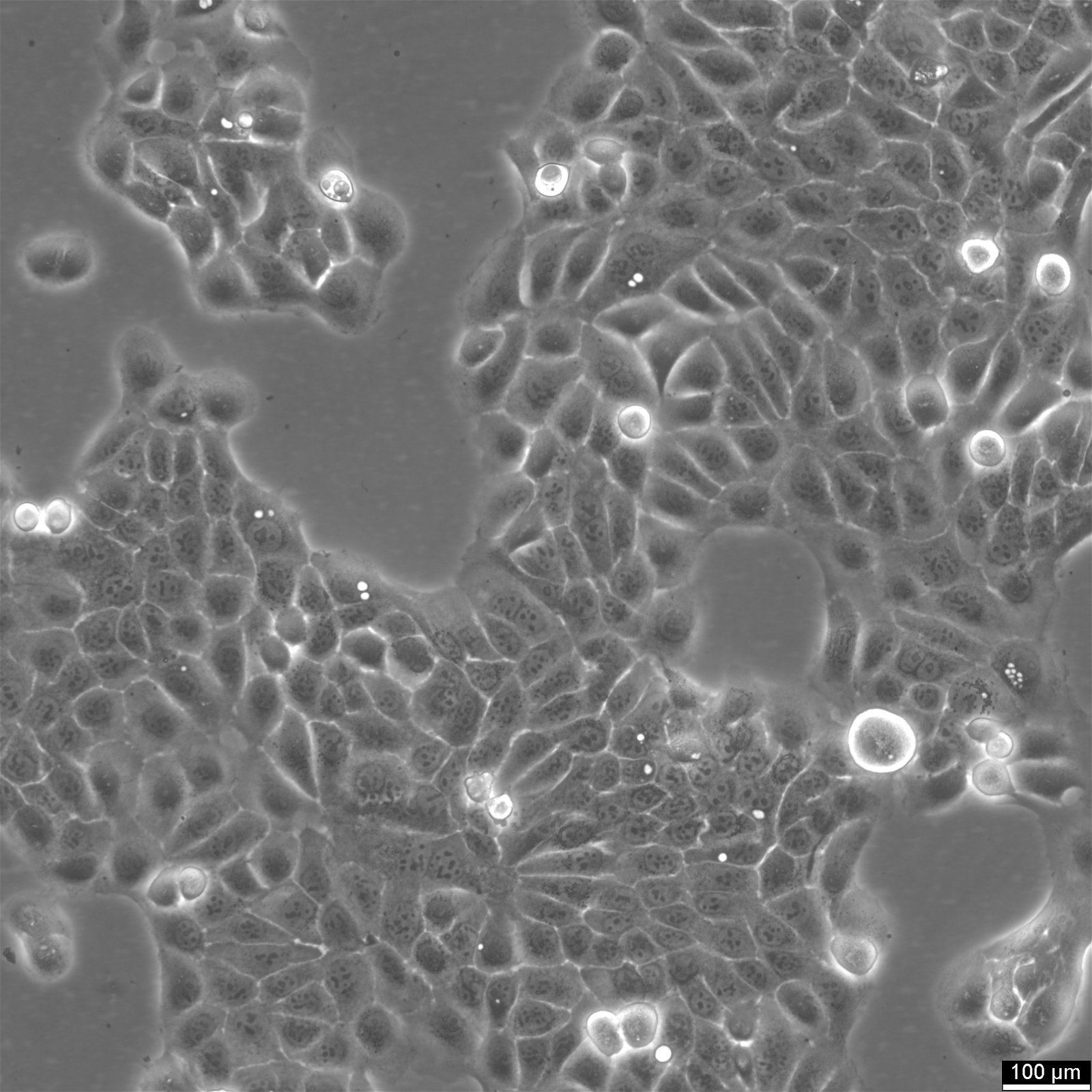
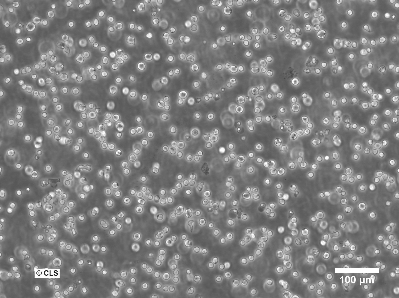
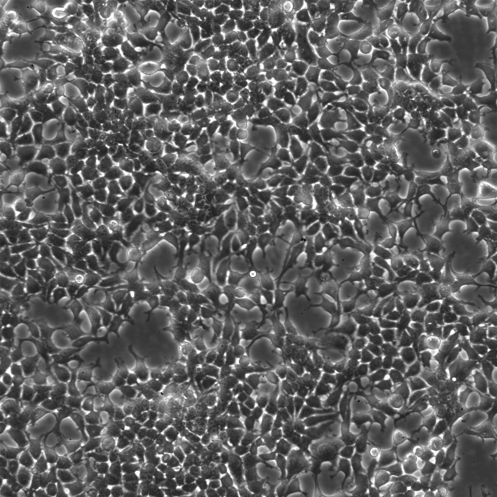
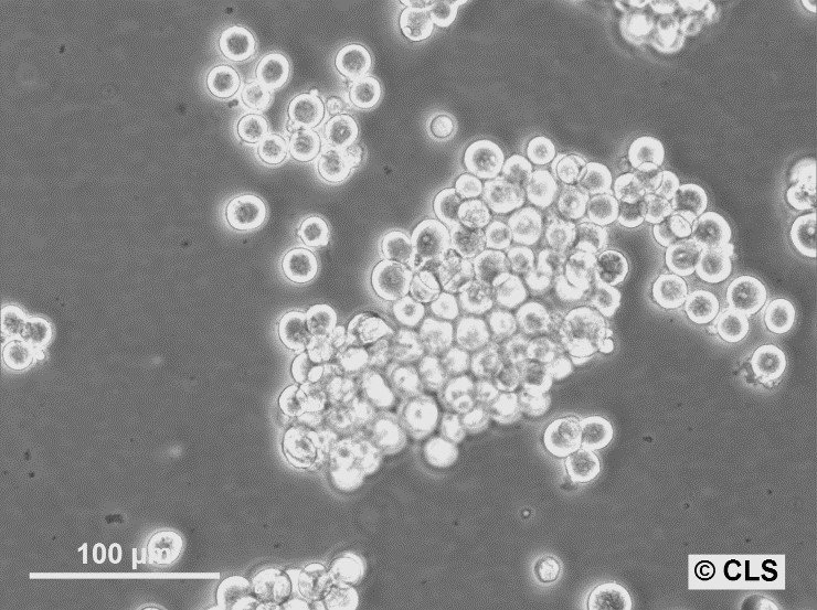
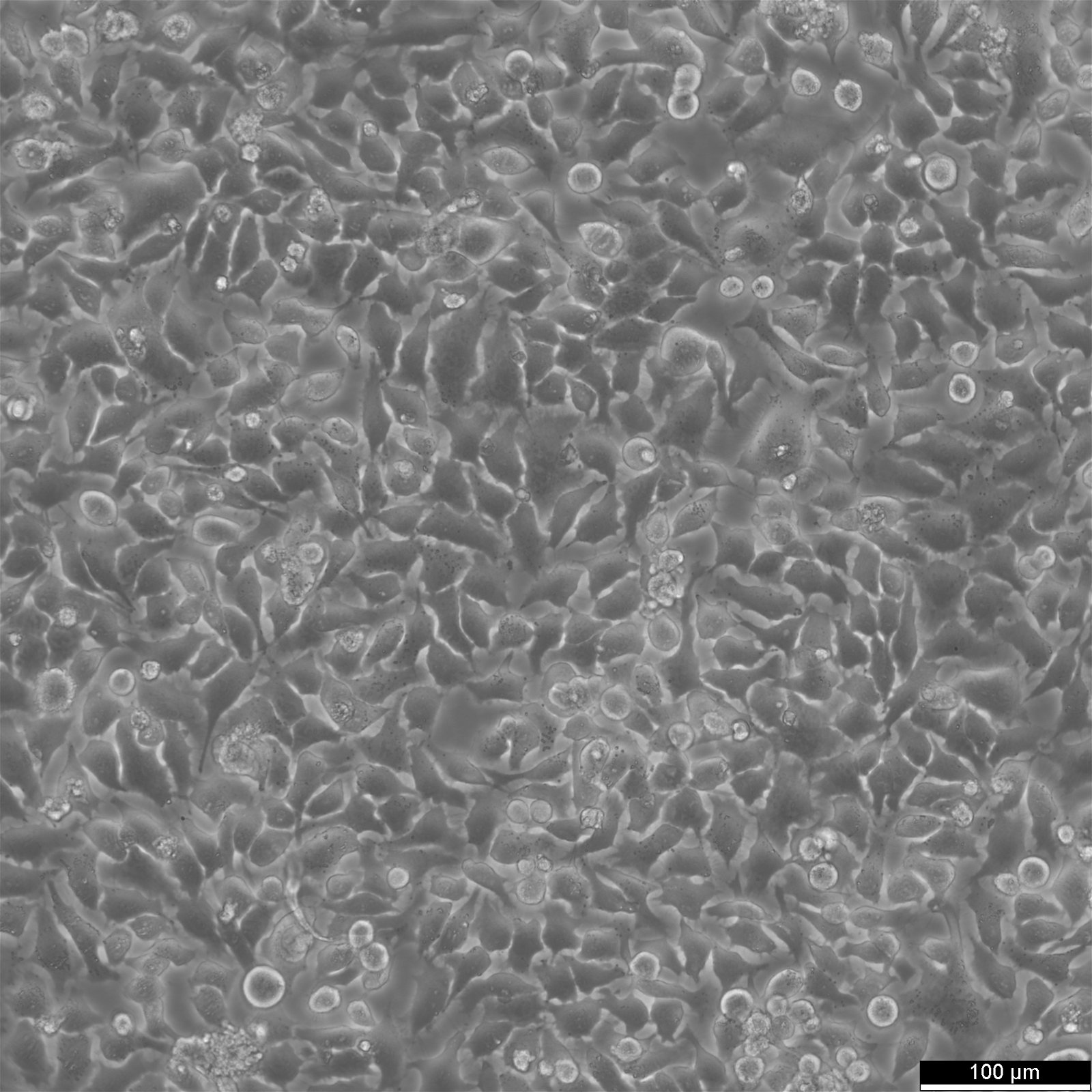
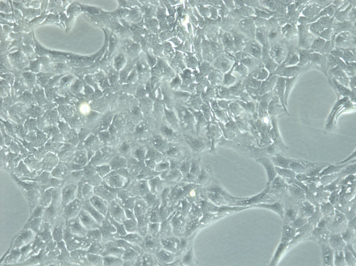
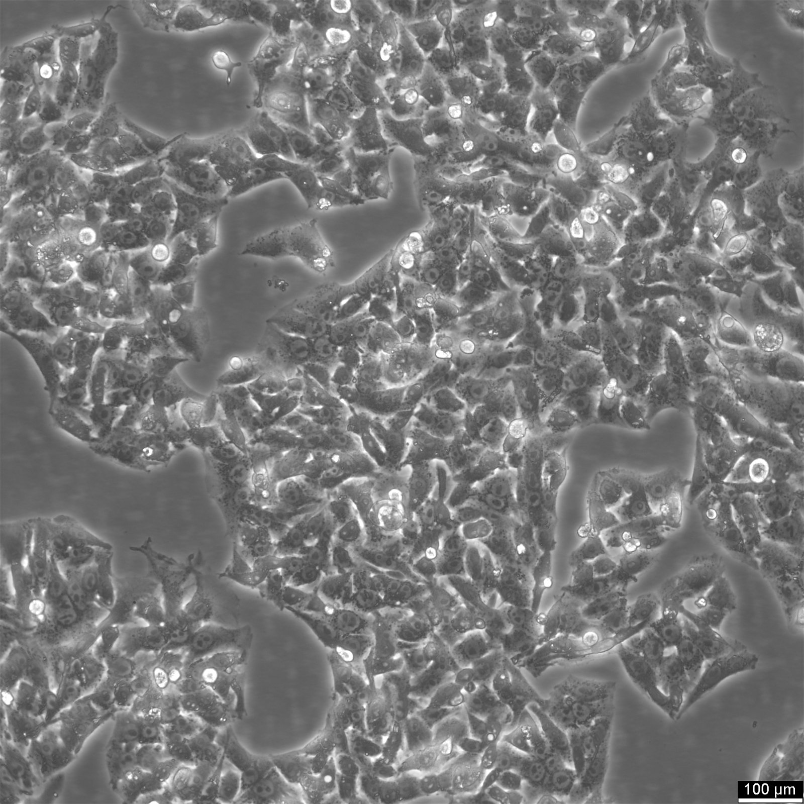
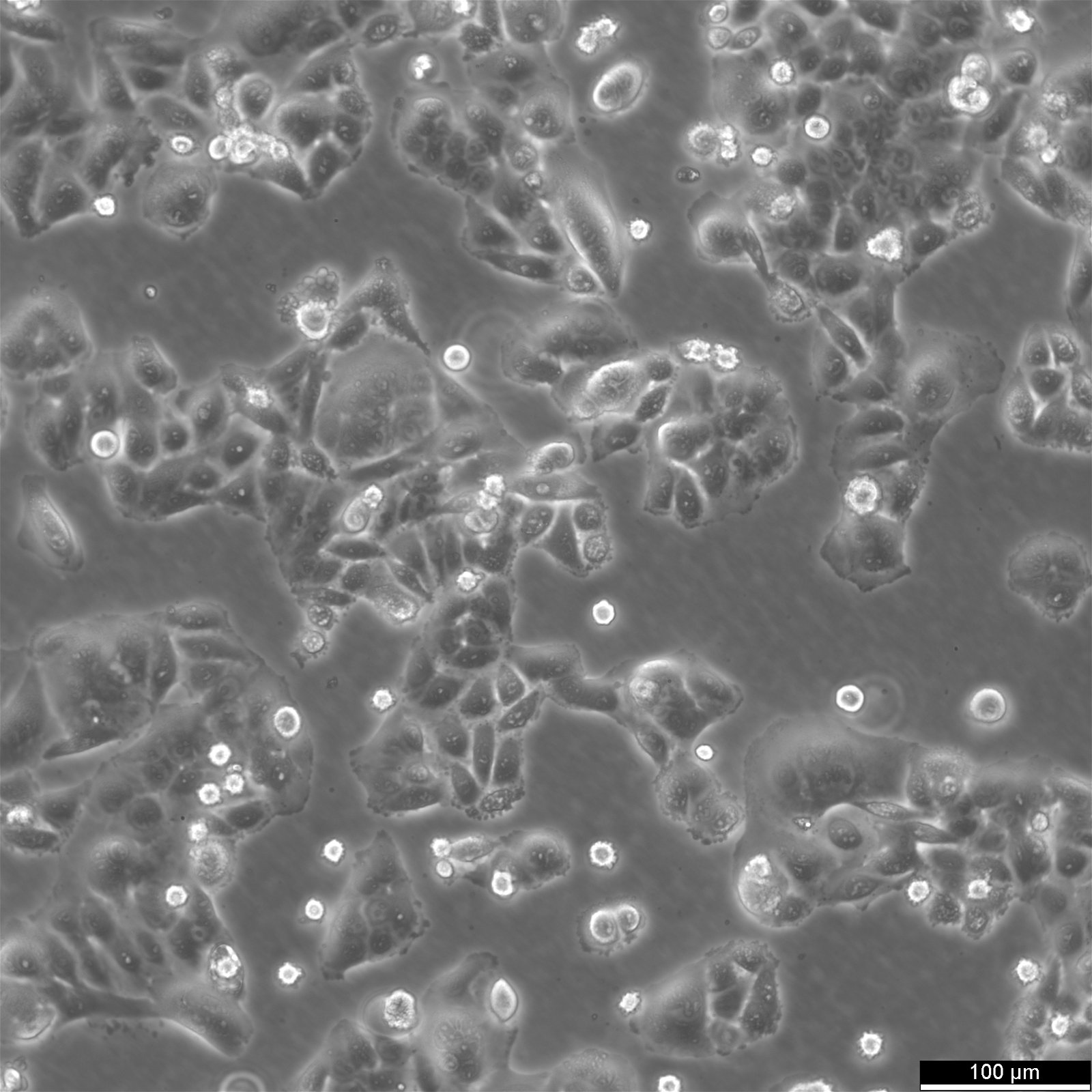
Menschliche Zellen
Willkommen bei Cytion, Ihrer ersten Adresse für authentifizierte und kontaminationsfreie humane Zelllinien. Unsere umfangreiche Zelllinienbank wurde sorgfältig zusammengestellt, um Ihre wissenschaftliche Forschung mit Zuverlässigkeit und Präzision zu unterstützen. Bei Cytion haben Sie Zugang zu einer breiten Palette menschlicher Zelllinien, die alle strengstens auf Reinheit und genetische Integrität geprüft wurden. Wir sind uns der entscheidenden Rolle bewusst, die qualitativ hochwertige Zellkulturen bei der Förderung der biomedizinischen Forschung spielen. Deshalb haben wir uns der Bereitstellung von Produkten verschrieben, auf die sich Forscher bei ihrer wichtigen Arbeit verlassen können.
Zellen für die Spitzenforschung
Entdecken Sie unser Portfolio an authentifizierten, validierten und mykoplasmenfreien Zelllinien, die sich für die biomedizinische Forschung, Proteinproduktion, Hybridomafusion, Virusvermehrung und vieles mehr eignen. Entscheiden Sie sich für Cytion, wenn es um die Bereitstellung von Zelllinien geht, wo jede Probe den Weg für Durchbrüche und Innovationen ebnet.
-
Produkte
-
Zellen und Zelllinien
-
Menschliche Zellen
- Brustkrebs-Zelllinien
- B-Lymphoblasten-Zelllinien
- Burkitt-Lymphom-Zelllinien
- Harnblasenkrebs-Zelllinien
- HaCaT-Zelllinien
- Hautkrebs-Zelllinien
- Hirntumor-Zelllinien
- Darmkrebs-Zelllinien
- Kopf-Hals-Krebs-Zelllinien
- Leberkrebs-Zelllinien
- Leukämie-Zelllinien
- Lungenkrebs-Zelllinien
- Knochenkrebs-Zelllinien
- Lymphom
- Magenkrebs-Zelllinien
- Myelom-Zelllinien
- Zelllinien für Nebennierenkrebs
- Neuroblastom-Zelllinien
- Zelllinien für Bauchspeicheldrüsenkrebs
- Prostatakrebs-Zelllinien
- Krebszelllinien des Reproduktionssystems
- Nierenkrebs-Zelllinien
- Zelllinien für Weichteilkrebs
- Hybride Zelllinien
- Spontan immortalisierte Zelllinien
- Transformierte Zelllinien
- Tierische Zellen
- Reporter-markierte Zellen
-
Menschliche Zellen
- Menschliche Primärzellen
- Stammzellen
- Medien und Reagenzien
- Nukleinsäuren
-
Zellen und Zelllinien
- Dienstleistung
- Wissenszentrum
- Übergang zu Cytion
| Organism | Menschen |
|---|---|
| Tissue | Das Ursprungsgewebe ist peripheres Blut |
| Disease | Leukämie |
| Organism | Menschen |
|---|---|
| Tissue | Haut |
| Disease | Melanom |
| Organism | Menschen |
|---|---|
| Tissue | Leber |
| Disease | Hepatozelluläres Karzinom |
| Organism | Menschen |
|---|---|
| Tissue | Eierstock |
| Disease | Hochgradiges seröses Adenokarzinom der Eierstöcke |
| Organism | Menschen |
|---|---|
| Tissue | Bauchspeicheldrüse |
| Disease | Duktales Adenokarzinom der Bauchspeicheldrüse |
| Organism | Menschen |
|---|---|
| Tissue | Doppelpunkt |
| Disease | Adenokarzinom |
| Organism | Menschen |
|---|---|
| Tissue | Niere |
| Organism | Menschen |
|---|---|
| Tissue | Knochen |
| Disease | Osteosarkom |
| Organism | Menschen |
|---|---|
| Tissue | Haut |
| Disease | Melanom |
| Organism | Menschen |
|---|---|
| Tissue | Lunge |
| Disease | Karzinom |
| Organism | Menschen |
|---|---|
| Tissue | Gebärmutterhals |
| Disease | Adenokarzinom |
| Organism | Menschen |
|---|---|
| Tissue | Eierstock |
| Disease | Seröses Zystadenokarzinom |
| Organism | Menschen |
|---|---|
| Tissue | Leber |
| Disease | Hepatozelluläres Karzinom |
| Organism | Menschen |
|---|---|
| Tissue | Doppelpunkt |
| Disease | Adenokarzinom, Grad IV, Dukes' Typ B. |
| Organism | Menschen |
|---|---|
| Tissue | Eierstock |
| Disease | Niedriggradiges seröses Ovarialkarzinom |
| Organism | Menschen |
|---|---|
| Tissue | Niere |
| Disease | Wilms-Tumor |
Übersicht über menschliche Zelllinien
Ganz gleich, ob Sie die Grundlagen der Krebsbiologie erforschen oder therapeutische Maßnahmen entwickeln, unsere Zelllinien bieten eine zuverlässige Grundlage für Ihre Forschungsarbeit und ebnen den Weg für Entdeckungen und Innovationen.
Unsere Sammlung wurde für zuverlässige, konsistente Forschungsergebnisse zusammengestellt. Vertrauen Sie auf die authentifizierten Zelllinien von Cytion, die strenge Qualitätsstandards erfüllen, frei von Krankheitserregern und identitätsgeprüft sind, damit Sie sich voll und ganz auf Ihre Forschung konzentrieren können.
Entdecken Sie unsere umfangreiche Auswahl, die mehr als 600 menschliche Krebszelllinien umfasst, die sorgfältig nach Krebsarten kategorisiert sind, um Ihren Such- und Auswahlprozess für einen effizienten Forschungsfortschritt zu vereinfachen.
Verständnis der Grundlagen von Zelllinien
Zellen, die immortalisiert und in vitro aus primären Explantaten von menschlichem Gewebe oder Körperflüssigkeit gezüchtet wurden, werden als humane Zelllinien bezeichnet.
Seit Anfang des 20. Jahrhunderts haben Wissenschaftler Zelllinien verwendet, um Einblicke in die Zellbiologie und den Stoffwechsel zu gewinnen. Zelllinien oder unsterbliche Zelllinien sind zu einem beliebten Modell in der Zellkulturliteratur geworden und dienen als gut charakterisierte und optimierte Einheit für pharmakologische Untersuchungen, biochemische Tests, die Synthese bioaktiver Substanzen usw. Kostengünstig, benutzerfreundlich und in der Lage, mehr Durchgänge zu durchlaufen als Primärzellen, werden Zelllinien von Wissenschaftlern bevorzugt. Zelllinien sind einfach zu manipulieren und zu vermehren, so dass sie für zahlreiche Screenings bevorzugt werden, da sie einen unbegrenzten Vorrat an Material bieten.
Immortalisierung von menschlichen Zelllinien
Immortalisierte Zellen können für immer kultiviert werden, wenn ihr Wachstum künstlich stimuliert wurde. Verschiedene Krebsarten und andere Zellen mit Chromosomenstörungen oder Mutationen, die eine unbegrenzte Vermehrung ermöglichen, bilden die Grundlage für immortalisierte Zelllinien.
Infolge ihrer schnellen Vermehrung wird die Schale oder der Kolben, in dem sich die immortalisierten Zellen befinden, überfüllt. Deshalb schaffen die Wissenschaftler mehr Platz für die sich vermehrenden Zellen, indem sie sie auf neue Platten übertragen (oder teilen).
Unterschiede zu Krebszelllinien
Es ist wichtig zu beachten, dass es einen grundlegenden Unterschied zwischen Tumorzellen und immortalisierten Zellen gibt: Tumorzellen weisen viele klassische Merkmale auf, wie z. B. Verlust der Kontakthemmung, schlechte Adhäsion und Apoptosehemmung, während immortalisierte Zellen ihren normalen Genotyp und Phänotyp beibehalten.
Methoden zur Erzeugung immortalisierter Zellen
Spontane Mutation
Während des Prozesses der Zellteilung und -vermehrung können bestimmte Ausgangszellen verändert werden und ihre Lebensdauer überschreiten. Diese Zellen werden für eine erweiterte Zellkultur geerntet und durchlaufen eine spontane Mutation, um zu Immortalisierungszellen zu werden. In den meisten Fällen verwandeln sich die Zellen jedoch in Tumorzellen, wodurch diese Technik unwirksam wird. Daher sind Tumorzellen das beste Beispiel für spontan immortalisierte Zellen, die genetische Veränderungen erworben haben, um die Seneszenz zu überleben und unsterblich zu werden.
Induktion der Immortalisierung von Zellen durch Virusgene
Zahlreiche virale Gene sind in der Lage, den Zellzyklus zu beeinflussen und so die Immortalisierung zu erreichen, indem sie die biologischen Bremsen der Proliferationsregulation ausschalten. Eine Möglichkeit, die Immortalisierung zu fördern, ist das T-Antigen des Simian-Virus 40 (SV40). Es hat sich gezeigt, dass das SV40-T-Antigen das einfachste und zuverlässigste Mittel für die Immortalisierung verschiedener Zelltypen ist, und sein Mechanismus bei der Immortalisierung von Zellen ist gut bekannt. Ein Beispiel ist der Zelltyp HEK293T (auch bekannt als 293T).
Telomerase Reverse Transkriptase (TERT) Proteinexpression
Telomerase ist ein Ribonukleoprotein, das die DNA-Sequenz der Telomere verlängern kann, wodurch die zelluläre Seneszenz verhindert wird und die Zellen sich unbegrenzt teilen können. Dieses Protein ist in den meisten somatischen Zellen inaktiv, aber wenn TERT exogen produziert wird, sind die Zellen in der Lage, genügend Telomerlängen aufrechtzuerhalten, um replikative Seneszenz zu verhindern. Derzeit ist die menschliche Telomerase-Reverse-Transkriptase (hTERT) die am häufigsten verwendete Methode zur Immortalisierung von Zellen.
Menschliche Zelllinien in biopharmazeutischen Anwendungen
Zelllinien werden nicht nur für die Modellierung biologischer Systeme und Krankheiten verwendet, sondern auch für praktische biotechnologische Zwecke bei der Herstellung von Proteinen, Viren und mehr. Entdecken Sie die für diese Anwendungen verwendeten Zellen:
Herstellung von rekombinanten Proteinen in Säugetier- und Insektenzellen
Aufgrund ihrer Fähigkeit zur Proteinsynthese sind eukaryotische Zelllinien für die Herstellung rekombinanter Proteine unverzichtbar geworden. Ihre Fähigkeit, die Proteinfaltung und den molekularen Zusammenbau zu erleichtern, übertrifft die anderer Systeme. Die Entwicklung von Expressionsvektoren und die Transfektion in das Wirtssystem sind die ersten Schritte bei der Herstellung rekombinanter Proteine, gefolgt von Zellauswahl, Klonierung, Screening und Bewertung. Um Qualitäts- und Skalierbarkeitskriterien zu erfüllen, benötigen die Hersteller rekombinanter Proteine effiziente und kostengünstige Expressionswirte.
Kultivierung von Viren
Die Einführung von Zellkulturmethoden hat die Isolierung und Vermehrung von Viren im Labor drastisch verändert. Für die Isolierung, den Nachweis und die Identifizierung von Viren bieten zellbasierte Produktionsverfahren eine praktische und kostengünstige Methode. Eine bessere Prozesskontrolle führt zu einem zuverlässigeren und besser charakterisierten Produkt mit schnelleren und kürzeren Produktionszyklen als bei Systemen auf Tierbasis oder Eibasis.
Wichtig sind zellbasierte Herstellungsverfahren für die Virenkultur und die Impfstoffherstellung für:
- Virusnachweis/-identifizierung
- Erforschung von Wirt-Pathogen-Interaktionen
- Virale Struktur und Replikation
- Impfstoffherstellung
Die Technologie der Hybridomazellen
Die Herstellung von monoklonalen Antikörpern, die für ein bestimmtes Antigen spezifisch sind, ist ein Bestandteil der Hybridomtechnologie. Durch die somatische Fusion von B-Lymphozyten der Milz mit unsterblichen Myelomzellen entsteht eine Hybridom-Zelllinie, die dauerhaft vermehrt werden kann, um klonal identische Antikörper zu produzieren, da diese Hybridomzellen die unbegrenzten Wachstumseigenschaften von Myelomzellen und die Fähigkeit zur Antikörpersekretion von B-Lymphozyten erben. Antikörper, die aus einer einzigen Hybridom-Zelllinie erzeugt werden, sind homogen und erkennen ein einziges Epitop auf einem Antigen.
Mithilfe der Hybridomtechnologie werden monoklonale Antikörper in den folgenden Bereichen eingesetzt:
- Biochemische Analyse: Monoklonale Antikörper verändern die Labordiagnostik. Biochemische Analysen (RIA, ELISA), Immunhistopathologie und diagnostische Bildgebung verwenden regelmäßig Antikörper (Immunszintigraphie).
- Immuntherapie: Menschliche, humanisierte und chimäre monoklonale Antikörper werden in der Immuntherapie zur Behandlung von Krebs, Autoimmunerkrankungen, Infektionskrankheiten, kardiovaskulären und anderen nicht-onkologischen Erkrankungen, als Adjuvans bei Organspenden und zur gezielten Verabreichung von Medikamenten eingesetzt.
- Proteinreinigung: Monoklonale Antikörper werden zur Reinigung von Proteinen eingesetzt und sind besonders vorteilhaft für die Reinigung rekombinanter Proteine (Immunaffinitätschromatographie).
Die Vorteile von menschlichen Zelllinien
- Konsistenz und Reproduzierbarkeit: Menschliche Zelllinien sind gut definiert und einheitlich, was zu konsistenten und reproduzierbaren Ergebnissen beiträgt.
- Leichte Kultivierung: Einfacher zu kultivieren als Primärzellen, da keine Gewebeentnahme erforderlich ist.
- Hohe Proteinproduktion: Kann große Proteinmengen für Assays produzieren.
- Genetische Modifikation: Kann so verändert werden, dass bestimmte Gene exprimiert werden, was für die Forschung nützlich ist.
Die Nachteile der Verwendung menschlicher Zelllinien
- Begrenzte Repräsentativität: Repräsentiert möglicherweise nicht genau die normalen In-vivo-Zellbedingungen.
- Genetische Drift: Im Laufe der Zeit kann es zu einem genetischen Drift kommen, der die Zelleigenschaften verändert.
- Veränderung im Laufe der Zeit: Eine längere Passage kann zum Verlust der ursprünglichen Zelleigenschaften führen.
- Reduzierte physiologische Relevanz: Die physiologische Relevanz für den Menschen kann eingeschränkt sein.
- Notwendigkeit der Validierung: Erfordert eine sorgfältige Validierung, um Authentizität und Reinheit zu gewährleisten.
Zukunft und Perspektiven
Seit der Etablierung der HeLa-Zelllinie wurden unsterbliche Krebszellen ausgiebig als biologische Modelle untersucht, um die Biologie des Krebses zu erforschen (einschließlich der Krebsentstehung, des Fortschreitens, der Metastasierung, der Mikroumgebung des Tumors und der Krebsstammzellen) und um neue Krebsmedikamente oder alternative Therapieformen wie die hyperthermische Therapie und den Einsatz von Nanopartikeln zu entwickeln. Aufgrund der Heterogenität von Krebs und arzneimittelresistenten Tumoren bei Patienten deuten jedoch zahlreiche Daten aus der Untersuchung von unsterblichen Krebszelllinien darauf hin, dass Krebszelllinien nicht repräsentativ genug sind. Die Forschung mit Krebszelllinien bietet die Chance, die Biologie von Tumoren besser zu verstehen und ermöglicht ein Hochdurchsatz-Screening für die Entwicklung von Medikamenten. Obwohl mehrere bedeutende Experimente mit Krebszelllinien durchgeführt wurden, liefern die Ergebnisse nur eine begrenzte Menge an Informationen und weisen eine schlechte klinische Korrelation auf. Dies ist einer der Gründe, warum diese Art von Studien die klinische Situation nicht vollständig abbildet. Daher können primäre Tumorzellkulturen (z. B. eine dreidimensionale Tumorzellkultur, die aus soliden Tumorproben gewonnen wird) genauere Informationen über bestimmte Krebsfälle liefern und die Entwicklung von therapeutischen Maßnahmen ermöglichen.

